- Home
- Medical news & Guidelines
- Anesthesiology
- Cardiology and CTVS
- Critical Care
- Dentistry
- Dermatology
- Diabetes and Endocrinology
- ENT
- Gastroenterology
- Medicine
- Nephrology
- Neurology
- Obstretics-Gynaecology
- Oncology
- Ophthalmology
- Orthopaedics
- Pediatrics-Neonatology
- Psychiatry
- Pulmonology
- Radiology
- Surgery
- Urology
- Laboratory Medicine
- Diet
- Nursing
- Paramedical
- Physiotherapy
- Health news
- Fact Check
- Bone Health Fact Check
- Brain Health Fact Check
- Cancer Related Fact Check
- Child Care Fact Check
- Dental and oral health fact check
- Diabetes and metabolic health fact check
- Diet and Nutrition Fact Check
- Eye and ENT Care Fact Check
- Fitness fact check
- Gut health fact check
- Heart health fact check
- Kidney health fact check
- Medical education fact check
- Men's health fact check
- Respiratory fact check
- Skin and hair care fact check
- Vaccine and Immunization fact check
- Women's health fact check
- AYUSH
- State News
- Andaman and Nicobar Islands
- Andhra Pradesh
- Arunachal Pradesh
- Assam
- Bihar
- Chandigarh
- Chattisgarh
- Dadra and Nagar Haveli
- Daman and Diu
- Delhi
- Goa
- Gujarat
- Haryana
- Himachal Pradesh
- Jammu & Kashmir
- Jharkhand
- Karnataka
- Kerala
- Ladakh
- Lakshadweep
- Madhya Pradesh
- Maharashtra
- Manipur
- Meghalaya
- Mizoram
- Nagaland
- Odisha
- Puducherry
- Punjab
- Rajasthan
- Sikkim
- Tamil Nadu
- Telangana
- Tripura
- Uttar Pradesh
- Uttrakhand
- West Bengal
- Medical Education
- Industry
Tips to prevent puncturing subclavian artery during IJV cannulation

The placement of a central venous catheter (CVC) has a 17.9% complication risk. Ultrasound guiding has been recommended as the gold standard of treatment to prevent problems and increase success rates. Ultrasound-guided cannulation, on the other hand, does not completely eliminate the risk of unintentional artery puncture. In a recent case report, the authors discussed how to prevent puncturing the subclavian artery during IJC cannulation.
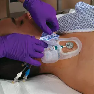
A 3-year-old female kid weighing 16.9 kg with a midline brain stem tumour and hydrocephalus was posted for elective tumour removal in this case report. On the first attempt, the right IJV was pierced with the needle angled at a 45° angle to insert a CVC. Following blood aspiration, a guide wire was successfully fed through the needle, dilated, and a 5.5 Fr paediatric multi-lumen CVC with blue flex tip was placed. Free blood aspiration was tested at all three ports. A sterile clear dressing was placed to the CVC at the 8 cm point. An arterial waveform was seen when the transducer was attached to the CVC. Ultrasound imaging was used to check the catheter's location in the artery before it was removed. The puncture site and IJV were located slightly to the right of the carotid artery, as predicted. A pulsing vessel underneath the jugular vein, which might be the subclavian artery, was discovered after further scanning and changing probe angles (SA). The catheter was shown to pass into the internal jugular canal and then lay in the SA, which was unexpected.
Ultrasound indicated that the SA was around 2.5 mm deeper than the IJV. The needle tip entering the SA might be due to indentation of the IJV wall when puncturing the vein or displacement of the needle tip after the probe was withdrawn. There are five crucial aspects to remember while cannulating IJV in young patients with the SA just a few millimetres distant.
• First, during piercing, the needle should be maintained angulated in regard to the skin, close to 30° rather than 45°, since this tangential approach leads in reduced posterior forces that tend to collapse the vein wall. When the IJV diameter is smaller than the needle's longitudinal length of bevel, angulation may aid.
• Second, double wall puncture should be remembered where the vein diameter is limited.
• Third- When the needle approaches the vein and puncture is performed, it is critical that the tip of the needle be continually recognised using ultrasound.
• Fourth, before dilatation of the vessel, ultrasonography should be used to validate the course of the guide wire.
• Finally, in youngsters, a higher neck approach may be used to extend the distance between the IJV and the SA, lowering the risk of unintentional puncture.
Reference –
Al Saadi, Nada RS; Haris, Aziz1; Khan, Rashid M1; Kaul, Naresh1, Iatrogenic catheterisation of subclavian artery while cannulating internal jugular vein, Indian Journal of Anaesthesia: April 2022 - Volume 66 - Issue 4 - p 304-305
doi: 10.4103/ija.ija_977_21
MBBS, MD (Anaesthesiology), FNB (Cardiac Anaesthesiology)
Dr Monish Raut is a practicing Cardiac Anesthesiologist. He completed his MBBS at Government Medical College, Nagpur, and pursued his MD in Anesthesiology at BJ Medical College, Pune. Further specializing in Cardiac Anesthesiology, Dr Raut earned his FNB in Cardiac Anesthesiology from Sir Ganga Ram Hospital, Delhi.
Dr Kamal Kant Kohli-MBBS, DTCD- a chest specialist with more than 30 years of practice and a flair for writing clinical articles, Dr Kamal Kant Kohli joined Medical Dialogues as a Chief Editor of Medical News. Besides writing articles, as an editor, he proofreads and verifies all the medical content published on Medical Dialogues including those coming from journals, studies,medical conferences,guidelines etc. Email: drkohli@medicaldialogues.in. Contact no. 011-43720751


