- Home
- Medical news & Guidelines
- Anesthesiology
- Cardiology and CTVS
- Critical Care
- Dentistry
- Dermatology
- Diabetes and Endocrinology
- ENT
- Gastroenterology
- Medicine
- Nephrology
- Neurology
- Obstretics-Gynaecology
- Oncology
- Ophthalmology
- Orthopaedics
- Pediatrics-Neonatology
- Psychiatry
- Pulmonology
- Radiology
- Surgery
- Urology
- Laboratory Medicine
- Diet
- Nursing
- Paramedical
- Physiotherapy
- Health news
- Fact Check
- Bone Health Fact Check
- Brain Health Fact Check
- Cancer Related Fact Check
- Child Care Fact Check
- Dental and oral health fact check
- Diabetes and metabolic health fact check
- Diet and Nutrition Fact Check
- Eye and ENT Care Fact Check
- Fitness fact check
- Gut health fact check
- Heart health fact check
- Kidney health fact check
- Medical education fact check
- Men's health fact check
- Respiratory fact check
- Skin and hair care fact check
- Vaccine and Immunization fact check
- Women's health fact check
- AYUSH
- State News
- Andaman and Nicobar Islands
- Andhra Pradesh
- Arunachal Pradesh
- Assam
- Bihar
- Chandigarh
- Chattisgarh
- Dadra and Nagar Haveli
- Daman and Diu
- Delhi
- Goa
- Gujarat
- Haryana
- Himachal Pradesh
- Jammu & Kashmir
- Jharkhand
- Karnataka
- Kerala
- Ladakh
- Lakshadweep
- Madhya Pradesh
- Maharashtra
- Manipur
- Meghalaya
- Mizoram
- Nagaland
- Odisha
- Puducherry
- Punjab
- Rajasthan
- Sikkim
- Tamil Nadu
- Telangana
- Tripura
- Uttar Pradesh
- Uttrakhand
- West Bengal
- Medical Education
- Industry
Cone-beam CT images may help identify several risk indicators of midfacial gingival recession in esthetic region

Cone-beam CT images may help identify several risk indicators of midfacial gingival recession in esthetic regions suggests a new study published in the Journal of Periodontology.
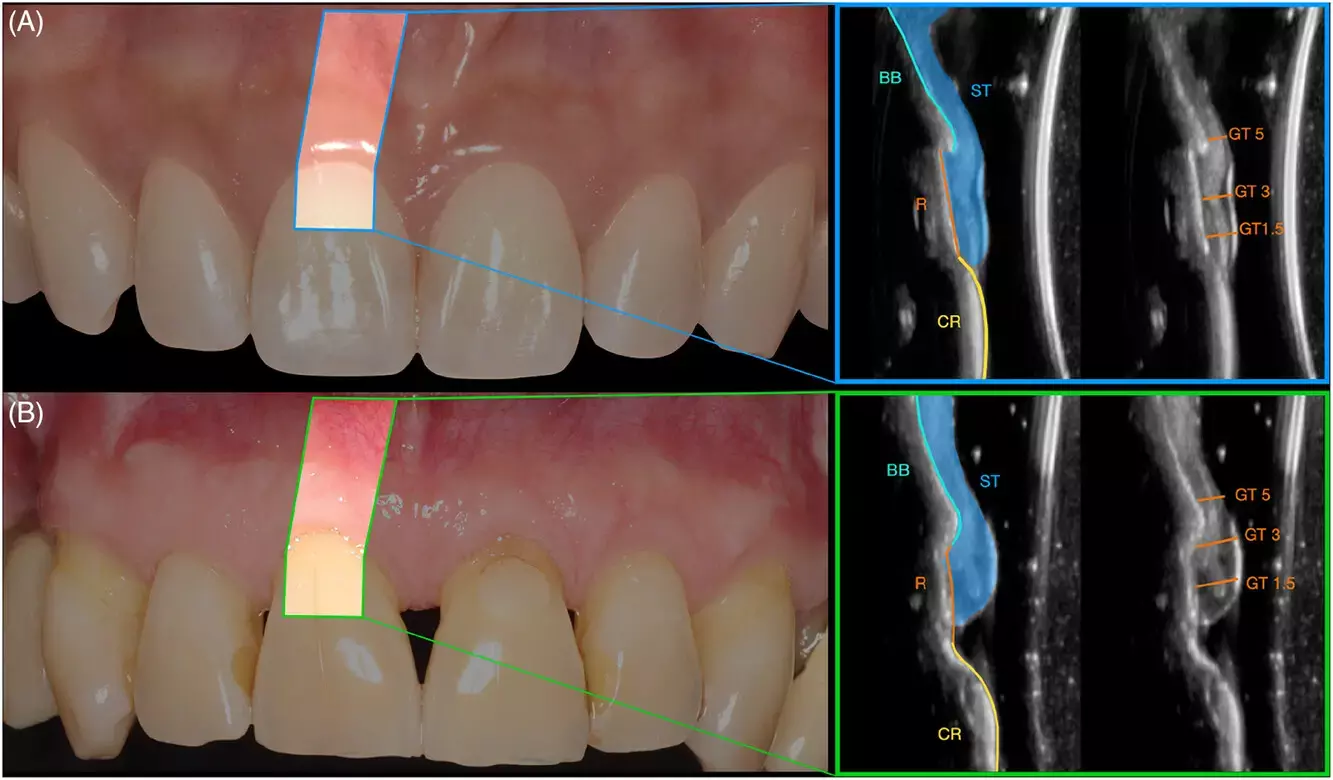
A study was done to evaluate the risk indicators associated with midfacial gingival recessions (GR) in the natural dentition esthetic regions. Cone-beam computed tomography (CBCT) results of thirty-seven subjects presenting with 268 eligible teeth were included in the cross-sectional study. Clinical measurements included the presence/absence of midfacial GR; the depth of the midfacial, mesial, and distal gingival recession; the recession type (RT); keratinized tissue width (KT); and attached gingiva width (AG). Questionnaires were utilized to capture patient-reported esthetics and dental hypersensitivity for each study tooth. Buccal bone dehiscence (cBBD) and buccal bone thickness (cBBT) were measured on the CBCT scans. High-frequency ultrasonography was performed to assess gingival thickness (GT) and buccal bone dehiscence (uBBD). Intraoral optical scanning was obtained to quantify the buccolingual position of each study site (3D profile analysis). Multilevel logistic regression analyses with generalized estimation equations were performed to assess the factors associated with the conditions of interest.
Results: The presence of midfacial GR was significantly associated with the history of periodontal treatment for pocket reduction (OR 7.99, p = 0.006), KT (OR 0.62, p < 0.001), cBBD (OR 2.30, p = 0.015), GT 1.5 mm from the gingival margin (OR 0.18, p = 0.04) and 3D profile 1 mm from the gingival margin (OR 1.04, p = 0.001). The depth of midfacial GR was significantly correlated to previous history of periodontal treatment (OR 0.96, p = 0.001), KT (OR −0.18, p < 0.001), presence of bone fenestration (OR 0.24, p = 0.044), and cBBD (OR 0.43, p < 0.001). The depth of midfacial GR was also the only factor associated with patient-reported esthetics (OR −3.38, p = 0.022), while KT (OR 0.77, p = 0.018) and AG (OR 0.82, p = 0.047) were significantly correlated with patient-reported dental hypersensitivity. Several risk indicators of midfacial and interproximal GR in the esthetic region were identified. The use of imaging technologies allowed for detection of parameters associated with the conditions of interest, and, therefore, their incorporation in future clinical studies is advocated. Ultrasonography could be preferred over CBCT for a noninvasive assessment of periodontal phenotype.
Reference:
Mascardo KC, Tomack J, Chen C-Y, et al. Risk indicators for gingival recession in the esthetic zone: A cross-sectional clinical, tomographic, and ultrasonographic study. J Periodontol. 2024; 1-12. https://doi.org/10.1002/JPER.23-0357
Keywords:
Cone-beam CT images, risk indicators, midfacial gingival recession, recession in gingiva, recession in gums esthetic region, Mascardo KC, Tomack J, Chen C-Y, Journal of Periodontology
Dr. Shravani Dali has completed her BDS from Pravara institute of medical sciences, loni. Following which she extensively worked in the healthcare sector for 2+ years. She has been actively involved in writing blogs in field of health and wellness. Currently she is pursuing her Masters of public health-health administration from Tata institute of social sciences. She can be contacted at editorial@medicaldialogues.in.
Dr Kamal Kant Kohli-MBBS, DTCD- a chest specialist with more than 30 years of practice and a flair for writing clinical articles, Dr Kamal Kant Kohli joined Medical Dialogues as a Chief Editor of Medical News. Besides writing articles, as an editor, he proofreads and verifies all the medical content published on Medical Dialogues including those coming from journals, studies,medical conferences,guidelines etc. Email: drkohli@medicaldialogues.in. Contact no. 011-43720751


