- Home
- Medical news & Guidelines
- Anesthesiology
- Cardiology and CTVS
- Critical Care
- Dentistry
- Dermatology
- Diabetes and Endocrinology
- ENT
- Gastroenterology
- Medicine
- Nephrology
- Neurology
- Obstretics-Gynaecology
- Oncology
- Ophthalmology
- Orthopaedics
- Pediatrics-Neonatology
- Psychiatry
- Pulmonology
- Radiology
- Surgery
- Urology
- Laboratory Medicine
- Diet
- Nursing
- Paramedical
- Physiotherapy
- Health news
- Fact Check
- Bone Health Fact Check
- Brain Health Fact Check
- Cancer Related Fact Check
- Child Care Fact Check
- Dental and oral health fact check
- Diabetes and metabolic health fact check
- Diet and Nutrition Fact Check
- Eye and ENT Care Fact Check
- Fitness fact check
- Gut health fact check
- Heart health fact check
- Kidney health fact check
- Medical education fact check
- Men's health fact check
- Respiratory fact check
- Skin and hair care fact check
- Vaccine and Immunization fact check
- Women's health fact check
- AYUSH
- State News
- Andaman and Nicobar Islands
- Andhra Pradesh
- Arunachal Pradesh
- Assam
- Bihar
- Chandigarh
- Chattisgarh
- Dadra and Nagar Haveli
- Daman and Diu
- Delhi
- Goa
- Gujarat
- Haryana
- Himachal Pradesh
- Jammu & Kashmir
- Jharkhand
- Karnataka
- Kerala
- Ladakh
- Lakshadweep
- Madhya Pradesh
- Maharashtra
- Manipur
- Meghalaya
- Mizoram
- Nagaland
- Odisha
- Puducherry
- Punjab
- Rajasthan
- Sikkim
- Tamil Nadu
- Telangana
- Tripura
- Uttar Pradesh
- Uttrakhand
- West Bengal
- Medical Education
- Industry
Two-step intraoral scans of edentulous mandibular arch may significantly decrease distortion while taking denture definitive impressions

Two-step intraoral scans of edentulous mandibular arch may significantly decrease distortion while taking denture definitive impressions suggests a new study published in the Journal of prosthetic dentistry.
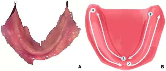
Manufacturers of several intraoral scanners have recommended a 2-step strategy for scanning the edentulous mandible. The 2-step technique requires scanning one side first and then moving to the other side. However, whether inconsistency in stitching occurs that results in loss of accuracy or distortion is unclear.
The purpose of this clinical study was to measure the potential distortion of intraoral scans of edentulous mandibular arches made with a 2-step scanning strategy and to assess their differences with conventional impressions.
Twenty mandibular edentulous arches were scanned by 1 investigator with an intraoral scanner using a 2-step scanning strategy, and a corresponding polysulfide conventional impression was obtained. The conventional impression was then immediately scanned with the same intraoral scanner. The obtained standard tessellation language (STL) files were superimposed with a surface-matching software program. After a preliminary alignment, the STL meshes were trimmed and reoriented; then, the final alignment was carried out and meshes moved to a metrology software program where their mean distance was measured. In addition, a surface curve (SIOS) was traced on the intraoral scan from the right to left retromolar pad along the residual ridge and automatically projected onto to the conventional impression scan to obtain a new curve (SC). The mean distance between SIOS and SC was measured and recorded as an indicator of the distortion by considering the X-, Y-, and Z-axes and the overall 3-dimensional (3D) deviation. The analysis was performed for the full curve length and after dividing it into 6 regions of interest. Univariate and multivariate statistical analyses were used to investigate the significance of the extent of the mean 3D distance, as well as the effects of measurement positions (side and region) between and within patients on differences along the X-, Y-, and Z-axes (α=.05).
Results
The mean (−0.08 mm; standard error: 0.025) 3D distance between the intraoral scan and conventional impression was significantly different from zero (P=.003). No significant effect of the factor “side” was found by using generalized estimated equation models for the X-, Y-, and Z-axes, and global 3D deviations between SIOS and SC (P>.05), which appeared to exclude distortion. Conversely, a significant effect was found for the factor “region” (P<.05), with no significant differences (P>.05) between corresponding regions on the 2 sides.
Intraoral scans of the edentulous mandibular arch made in a 2-step procedure did not exhibit significant distortion in comparison with conventional impressions.
Reference:
Assessment of distortion of intraoral scans of edentulous mandibular arch made with a 2-step scanning strategy: A clinical study. Lucio Lo Russo, Roberto Sorrentino, Fariba Esperouz, Fernando Zarone, Carlo Ercoli, Laura Guida. Published:November 03, 2023DOI:https://doi.org/10.1016/j.prosdent.2023.09.029
Keywords:
Two-step, intraoral, scans, edentulous, mandibular, arch, significantly, decrease, distortion, while, taking, denture, definitive, impressions, Lucio Lo Russo, Roberto Sorrentino, Fariba Esperouz, Fernando Zarone, Carlo Ercoli, Laura Guida, Journal of prosthetic dentistry
Dr. Shravani Dali has completed her BDS from Pravara institute of medical sciences, loni. Following which she extensively worked in the healthcare sector for 2+ years. She has been actively involved in writing blogs in field of health and wellness. Currently she is pursuing her Masters of public health-health administration from Tata institute of social sciences. She can be contacted at editorial@medicaldialogues.in.
Dr Kamal Kant Kohli-MBBS, DTCD- a chest specialist with more than 30 years of practice and a flair for writing clinical articles, Dr Kamal Kant Kohli joined Medical Dialogues as a Chief Editor of Medical News. Besides writing articles, as an editor, he proofreads and verifies all the medical content published on Medical Dialogues including those coming from journals, studies,medical conferences,guidelines etc. Email: drkohli@medicaldialogues.in. Contact no. 011-43720751


