- Home
- Medical news & Guidelines
- Anesthesiology
- Cardiology and CTVS
- Critical Care
- Dentistry
- Dermatology
- Diabetes and Endocrinology
- ENT
- Gastroenterology
- Medicine
- Nephrology
- Neurology
- Obstretics-Gynaecology
- Oncology
- Ophthalmology
- Orthopaedics
- Pediatrics-Neonatology
- Psychiatry
- Pulmonology
- Radiology
- Surgery
- Urology
- Laboratory Medicine
- Diet
- Nursing
- Paramedical
- Physiotherapy
- Health news
- Fact Check
- Bone Health Fact Check
- Brain Health Fact Check
- Cancer Related Fact Check
- Child Care Fact Check
- Dental and oral health fact check
- Diabetes and metabolic health fact check
- Diet and Nutrition Fact Check
- Eye and ENT Care Fact Check
- Fitness fact check
- Gut health fact check
- Heart health fact check
- Kidney health fact check
- Medical education fact check
- Men's health fact check
- Respiratory fact check
- Skin and hair care fact check
- Vaccine and Immunization fact check
- Women's health fact check
- AYUSH
- State News
- Andaman and Nicobar Islands
- Andhra Pradesh
- Arunachal Pradesh
- Assam
- Bihar
- Chandigarh
- Chattisgarh
- Dadra and Nagar Haveli
- Daman and Diu
- Delhi
- Goa
- Gujarat
- Haryana
- Himachal Pradesh
- Jammu & Kashmir
- Jharkhand
- Karnataka
- Kerala
- Ladakh
- Lakshadweep
- Madhya Pradesh
- Maharashtra
- Manipur
- Meghalaya
- Mizoram
- Nagaland
- Odisha
- Puducherry
- Punjab
- Rajasthan
- Sikkim
- Tamil Nadu
- Telangana
- Tripura
- Uttar Pradesh
- Uttrakhand
- West Bengal
- Medical Education
- Industry
Emerging Imaging Technologies for Parathyroid Gland Identification and Vascular Assessment in Thyroid Surgery

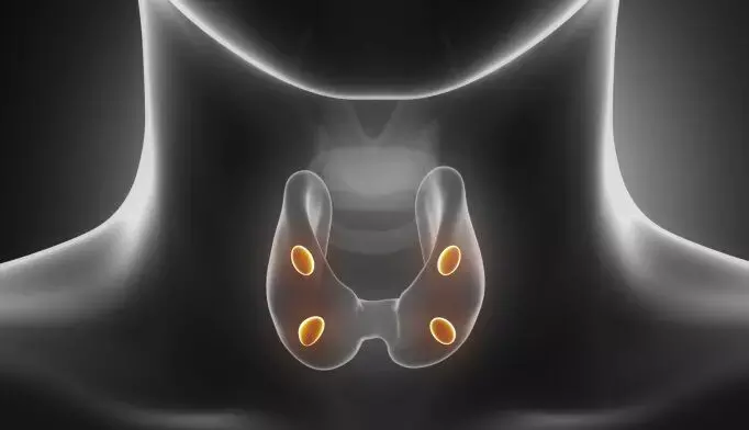
Hypoparathyroidism, an endocrine disorder characterized by low calcium and absent or insufficient circulating parathyroid hormone (PTH), is most commonly a consequence of surgical injury or removal of PGs during anterior neck surgery, less commonly autoimmune or genetic disorders. Depending on the definition of hypoparathyroidism, rates of permanent postoperative hypoparathyroidism, herein defined as failure of functional recovery 6 to 12 months after thyroidectomy, range from 4% to 12%, higher in patients undergoing neck dissection or partial or total thyroidectomy concurrent with total laryngectomy (especially after radiation therapy). Subcapsular surgical dissection is not always sufficient to preserve PGs and prevent the considerable morbidity, in some cases mortality, resulting from hypoparathyroidism caused by devascularization or inadvertent removal of PGs during surgery. Emerging imaging technologies hold promise to improve identification and preservation of PGs during thyroid surgery.
Amanda L. et al comprehensively reviewed PG identification and vascular assessment using near-infrared autofluorescence (NIRAF)—both label free and in combination with indocyanine green (ICG)— based on a comprehensive literature review and provide a manual for possible implementation of these emerging technologies in thyroid surgery.
This narrative review comprehensively reviews PG identification and vascular assessment using near-infrared autofluorescence (NIRAF)—both label free and in combination with indocyanine green—based on a comprehensive literature review and offers a manual for possible implementation these emerging technologies in thyroid surgery.
Despite surgical advances, preservation of PGs remains challenging due to their small size and variable location. The morbidity associated with hypoparathyroidism following thyroid surgery is substantial and includes decreased quality of life; kidney, neurologic, and musculoskeletal complications; and even increased mortality.
Management of permanent postoperative hypoparathyroidism can be challenging, often requiring calcium supplements and activated vitamin D, and in some patients magnesium, thiazide diuretics, phosphate binders, dietary/lifestyle changes, and recombinant human intact PTH. Undertreatment of pregnant or breastfeeding patients can lead to maternal hypocalcemia (linked to premature delivery, intrauterine fetal hyperparathyroidism, and fetal death), while overtreatment can result in abortion, stillbirth, perinatal death, and neonatal tetany. Postsurgical hypoparathyroidism with prior gastric bypass often results in recalcitrant hypocalcemia, at times requiring prolonged parenteral calcium, and even reversal of the gastric bypass with varying results. Leveraging surgical technologies to prevent PG injury is therefore important.
Parathyroid Gland Identification
- Comparison of Probe-Based and Camera-Based Technologies
Early PG localization may inform real-time surgical adjustments to minimize manipulation of PGs, thereby preventing injury. Commercially available NIR fluorescence systems for thyroid surgery can be divided into 2 groups: probe based and camera based. In 2018, US Food and Drug Administration clearance was granted to (1) probe-based PTeye (Medtronic) and (2) camerabased Fluobeam 800 and Fluobeam LX (Fluoptics) to serve as labelfree NIRAF detection devices to identify PGs intraoperatively.
Current systems have benefits and limitations, and each surgeon should consider personal outcomes, surgical volume, and cost. With experience and proper calibration, NIRAF technologies enable earlier and improved PG identification compared with the current gold standard, the surgeon’s unaided eye. The NIRAF imaging can identify 90% to 100% of PGs with 90% to 100% sensitivity and accuracy.
In one study, when compared concurrently in the same set of 20 patients, probe-based NIRAF was more sensitive in PG identification vs camera image– based NIRAF (detection rate of 97% and 91%, respectively). A meta-analysis of 2062 patients found a 98% sensitivity and 99% specificity of NIRAF (probe or camera based) to identify PG during thyroid surgery. A separate meta-analysis of 1198 patients found NIRAF can identify PGs with a sensitivity, specificity, negative predictive value, and positive predictive value of 97%, 92%, 95%, and 95%, respectively, with a higher diagnostic accuracy using probebased vs camera-based systems.
The NIRAF detection systems may be used to scan the surface of the surgical field and localize thinly covered PGs, much like a nerve monitor probe can identify the path of the recurrent laryngeal nerve. In 2 studies (n = 21016 and n = 3827), PG mapping with NIRAF imaging systems was possible in 37% to 92% of patients before PG exposure.
Importantly, NIRAF technology does not supplant sound judgment and surgical experience. There is variability in reports of whether the use of NIRAF reduces postoperative hypocalcemia, and its use has not been shown definitively to reduce permanent postthyroidectomy hypocalcemia. The learning curve of adoption of these nascent technologies requires study, including operator variability and extrinsic variables (such as ambient light), in addition to variables affecting PG autofluorescence intensity and interpretation.
Parathyroid Gland Vascularization
- Intraoperative Assessment of Parathyroid Perfusion
Several intraoperative techniques to evaluate PG perfusion during thyroidectomy have been studied. Provided perfusion is confirmatory of postoperative PG function, intraoperative preservation of PG perfusion could reduce rates of postoperative hypoparathyroidism.
ICG Fluorescence Imaging
The combination of NIR fluorescence imaging with ICG dye for realtime intraoperative evaluation of PG vascularization is gaining interest; NIRAF in conjunction with ICG imaging may provide information about perfusion and perhaps postoperative PG function. Indocyanine green injection during thyroidectomy can evaluate PG vascularity and perfusion, though ICG is not approved by the US Food andDrug Administration specifically for perfusion assessment in thyroid surgery. Intraoperative injection of ICG for PG localization has been compared with label-free NIRAF imaging with similar identification rates (95% vs 98%, respectively).
Several institutions have described early ICG injection after initial surgicalexposure. Once PGs have been identified, ICG fluorescence imaging can be used to assess PG vascular pedicles (realtime vascular mapping) and perfusion. Camera-based NIRAF detection methods and predissection ICG injection may provide a spatial guide of PG and associated vascular anatomy, thereby optimizing PG management. This technique requires correctly timing the ICG injection. The best images are obtained after the first injection due to remnant background ICG fluorescence during subsequent injections. Importantly, NIRAF cannot be detected after ICG administration because the background ICG fluorescence is more intense than the NIRAF.
The most common timing for intraoperative ICG injection is after completion of total thyroidectomy. The ICG injection, however, can occur at various times throughout the procedure, such as after initial PG dissection to aid in preservation of feeding vessels or after completion of the first side to aid in decision-making regarding extent of second-side dissection. Although not yet studied systematically, injection after completion of the first side may be appropriate in certain high-risk contexts (eg, prior gastric bypass) or reoperative cases. Used in these contexts, surgeons may elect to stage contralateral thyroidectomy.
Emerging technologies hold promise to improve PG identification and preservation during thyroidectomy, and an integrated system enabling early PG identification and prediction of postoperative PG function would be ideal but is not yet available; rate of use of NIRAF technology, specifically in thyroid surgery, is not available. Additional research is needed to clarify the variables affecting the degree of fluorescence in NIRAF, standardize NIRAF signal quantification, standardize the parameters of ICG injection (dosing, timing of injection, and signal quantification), enable this technology to predict postoperative PG function, and understand the adoption learning curve and effect on surgical training as well as the financial effect of these emerging technologies on near-term costs (ie, operating time, reduction/ elimination of frozen section) and longer-term costs (ie, medications, surveillance costs). Long-term outcomes of key quality metrics are needed, and adequately powered randomized clinical trials evaluating PG preservation will guide adoption.
Source: Amanda L. Silver Karcioglu, MD; Frédéric Triponez; JAMA Otolaryngol Head Neck Surg. doi:10.1001/jamaoto.2022.4421
Dr Ishan Kataria has done his MBBS from Medical College Bijapur and MS in Ophthalmology from Dr Vasant Rao Pawar Medical College, Nasik. Post completing MD, he pursuid Anterior Segment Fellowship from Sankara Eye Hospital and worked as a competent phaco and anterior segment consultant surgeon in a trust hospital in Bathinda for 2 years.He is currently pursuing Fellowship in Vitreo-Retina at Dr Sohan Singh Eye hospital Amritsar and is actively involved in various research activities under the guidance of the faculty.
Dr Kamal Kant Kohli-MBBS, DTCD- a chest specialist with more than 30 years of practice and a flair for writing clinical articles, Dr Kamal Kant Kohli joined Medical Dialogues as a Chief Editor of Medical News. Besides writing articles, as an editor, he proofreads and verifies all the medical content published on Medical Dialogues including those coming from journals, studies,medical conferences,guidelines etc. Email: drkohli@medicaldialogues.in. Contact no. 011-43720751


