- Home
- Medical news & Guidelines
- Anesthesiology
- Cardiology and CTVS
- Critical Care
- Dentistry
- Dermatology
- Diabetes and Endocrinology
- ENT
- Gastroenterology
- Medicine
- Nephrology
- Neurology
- Obstretics-Gynaecology
- Oncology
- Ophthalmology
- Orthopaedics
- Pediatrics-Neonatology
- Psychiatry
- Pulmonology
- Radiology
- Surgery
- Urology
- Laboratory Medicine
- Diet
- Nursing
- Paramedical
- Physiotherapy
- Health news
- Fact Check
- Bone Health Fact Check
- Brain Health Fact Check
- Cancer Related Fact Check
- Child Care Fact Check
- Dental and oral health fact check
- Diabetes and metabolic health fact check
- Diet and Nutrition Fact Check
- Eye and ENT Care Fact Check
- Fitness fact check
- Gut health fact check
- Heart health fact check
- Kidney health fact check
- Medical education fact check
- Men's health fact check
- Respiratory fact check
- Skin and hair care fact check
- Vaccine and Immunization fact check
- Women's health fact check
- AYUSH
- State News
- Andaman and Nicobar Islands
- Andhra Pradesh
- Arunachal Pradesh
- Assam
- Bihar
- Chandigarh
- Chattisgarh
- Dadra and Nagar Haveli
- Daman and Diu
- Delhi
- Goa
- Gujarat
- Haryana
- Himachal Pradesh
- Jammu & Kashmir
- Jharkhand
- Karnataka
- Kerala
- Ladakh
- Lakshadweep
- Madhya Pradesh
- Maharashtra
- Manipur
- Meghalaya
- Mizoram
- Nagaland
- Odisha
- Puducherry
- Punjab
- Rajasthan
- Sikkim
- Tamil Nadu
- Telangana
- Tripura
- Uttar Pradesh
- Uttrakhand
- West Bengal
- Medical Education
- Industry
Fortis, Mulund uses Nikaidohs Procedure to save life of 5-year old

Five-year old Sunita (name changed) was born with a complex heart condition.Her heart had a large hole between the two lower chambers.In medical terms, such condition is known as Ventricular Septal Defect or VSD. This congenital condition had the right lower chamber of her heart giving rise to both her great arteries. While the Ventricular Septal Defect in itself is not very common, Sunita's case was complicated as her Pulmonary Artery (the main artery extending to the lungs) was narrow and it arose in an abnormal position with respect to the Aorta (the main artery to the body). In medical terms, this condition is known as 'double outlet right ventricle with malposed great arteries with a large inlet Ventricular Septal Defect and severe Pulmonary Stenosis'.
One of the two children of a Palghar-resident, a fisherman;Sunitagrew up with the rare condition for five years, but the symptoms started to get more noticeable as she turned five. Her condition made her turn blue and she got tired easily with the slightest walking. Her lips and nails started turning purple. Even though her parents knew of the problem sinceshe was 3 month old, they were unable to help her. While money for the treatment might have been an issue, her parents were also nervousand very scared of the surgery that was suggested for her.
After avoiding treatment for over five years, Sunita's parents decided to explore options when the symptoms became even more visible and the child started feeling uneasy. Sunitaexperienced symptoms like shortness of breath, pallor, sweating while feeding etc. Upon the family doctor's advice, her parents approached Fortis Hospital, Mulund. Sunita's case was referred to Dr Vijay Agarwal, Head Paediatric Cardiac Surgery, Fortis Hospital,Mulund.
"Sunita's condition was rare due to the complications involved. Her Aorta was at wrong place - instead of being above the left ventricle, it was placed above the right ventricle. And this was further complicated by her narrowed Pulmonary Arteries. When we first assessed the patient in our OPD, we realized that we could approach Sunita's case intwo totally different ways.In the first approach, we could have performedtwo separate surgeries to fix two different conditions. Though this plan had low risk, the heart defects would not be corrected fully with this approach. The second option would be corrective, but radical, in which the heart would be opened up and the base of the Aorta would be cut off from the heart. The Aorta would then be re-stitched over the correct ventricle (left ventricle). The remaining steps would be routine.This surgery is called 'NikaidohProcedure'," informedDr Agarwal.
Surgeries for congenital heart disease have become common in the last few decades, but the certain complicated conditions – like Sunita's – are still a challenge to the congenital heart surgeons and very few surgeons across the world perform this surgery (Nikaidoh Operation). Yet, after deliberating the pros and cons of both the approaches, the team at Fortis Hospital, Mulund, decided in favour of the second option – NikaidohProcedure.
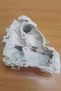
The team at Fortis Hospital, Mulund approached Sunita's case extremely carefully by eliminating any possibilities of error. The team first obtained a CT scan of the child's heart and with the help of the scans, a 3-Dimensional Print model of her heart was developed. The model gave the surgical team a clear idea about what to expect on the operating table. "With a precise3-D model of the heart, we were certain about the problem areas and were able to plan out the surgery with greater confidence of success," said DrSwati Garekar, Pediatric cardiologist, Fortis hospital, Mulund.
The first defect was solved through the NikaidohProcedure, where the Aorta was cut off from the right ventricle and was moved to above the left ventricle, which itself is a complex procedure as Aorta is the main artery of the body. "Given its complexity and risks involved, the NikaidohProcedure is rarely used in the city. But with our experience of handling similar cases in the past, the state-of-the-art facilities at Mulund's Fortis Hospital and with the help of 3D Print model of the heart, we were able to manage risks and performed the surgery successfully", she said.
Once the war of misplaced Aorta was won with the NikaidohProcedure, fixing the choked up Pulmonary arteries was a small battle. The second defect was solved with a conduit tube. "Many repair operations for congenital heart defects involve the replacement of valves and/or the insertion of Pulmonary Artery conduits to redirect blood flow," says Dr Agarwal. As second part of the procedure, Sunita's defective pulmonary arteries were removed as they were all choked up and were replaced with a conduit tube where one end of it was attached to right ventricle and the other end was attached to the lungs.
The surgery was carried out in January 2016 at Fortis Hospital, Mulund's Fortis Child Heart Mission. It lasted for 4 hours. Post-surgery, Sunita has recovered well. She is all set to be discharged home later this week. She is expected to have a good quality of life long term.


