- Home
- Medical news & Guidelines
- Anesthesiology
- Cardiology and CTVS
- Critical Care
- Dentistry
- Dermatology
- Diabetes and Endocrinology
- ENT
- Gastroenterology
- Medicine
- Nephrology
- Neurology
- Obstretics-Gynaecology
- Oncology
- Ophthalmology
- Orthopaedics
- Pediatrics-Neonatology
- Psychiatry
- Pulmonology
- Radiology
- Surgery
- Urology
- Laboratory Medicine
- Diet
- Nursing
- Paramedical
- Physiotherapy
- Health news
- Fact Check
- Bone Health Fact Check
- Brain Health Fact Check
- Cancer Related Fact Check
- Child Care Fact Check
- Dental and oral health fact check
- Diabetes and metabolic health fact check
- Diet and Nutrition Fact Check
- Eye and ENT Care Fact Check
- Fitness fact check
- Gut health fact check
- Heart health fact check
- Kidney health fact check
- Medical education fact check
- Men's health fact check
- Respiratory fact check
- Skin and hair care fact check
- Vaccine and Immunization fact check
- Women's health fact check
- AYUSH
- State News
- Andaman and Nicobar Islands
- Andhra Pradesh
- Arunachal Pradesh
- Assam
- Bihar
- Chandigarh
- Chattisgarh
- Dadra and Nagar Haveli
- Daman and Diu
- Delhi
- Goa
- Gujarat
- Haryana
- Himachal Pradesh
- Jammu & Kashmir
- Jharkhand
- Karnataka
- Kerala
- Ladakh
- Lakshadweep
- Madhya Pradesh
- Maharashtra
- Manipur
- Meghalaya
- Mizoram
- Nagaland
- Odisha
- Puducherry
- Punjab
- Rajasthan
- Sikkim
- Tamil Nadu
- Telangana
- Tripura
- Uttar Pradesh
- Uttrakhand
- West Bengal
- Medical Education
- Industry
Can Lamina Cribrosa Indicate Optic Neuritis in Multiple Sclerosis?

In a recent article published in Neurology India, Researchers in Turkey have found that the lamina cribrosa was thinner in Multiple Sclerosis patients with VEP pathology.
MS is a chronic, autoimmune, inflammatory demyelinating neurodegenerative disease of the central nervous system, which is usually characterised by variable clinical outcomes owing to recurrent episodes of immune-mediated demyelination, glial scar formation, and axonal loss. Optic neuritis (ON), an acute inflammatory demyelinating disorder of the optic nerve, is the first sign of MS in approximately 20% of MS patients. Approximately, half of MS patients develop ON during the course of the disease.
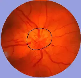
The intraocular part of the optic nerve is referred to as the optic nerve head; it is about 1 mm in length and is often described in terms of four parts: the lamina retinalis, lamina choroidalis, lamina cribrosa (LC), and retrolaminar layer. The LC consists of perforated hard connective tissue and elastic fibers. The glial tissue supporting the axons is most abundant in this layer. The LC is an essential web-like tissue layer, where abundant glia that support the axons of retinal ganglion cells are found; it allows the passage of nutrients and oxygen. Spectral domain optical coherence tomography (SD-OCT) is a noninvasive, safe, and easy imaging modality that is used to assess the thickness of retinal tissues and the retinal nerve fiber layer (RNFL) and the lamina cribrosa thickness (LCT).
The measurement of visual evoked potentials (VEPs) is the best way to reveal ON in MS patients. VEPs have been shown to detect ON even in silent cases that are clinically faint. However, although the specificity and sensitivity of VEPs are high in determining ON, they are not 100%. In spite recent advances in the treatment of MS, there is still no biomarker or method that can establish a definitive diagnosis. Although VEPs are currently considered the most sensitive and specific method for detecting ON in MS, abnormal VEP findings are detected in about 65% of MS patients without ON symptoms or history.
The authors divided the patients enrolled in this prospective, cross-sectional study into three groups. Group 1 consisted of 25 relapsing-remitting MS patients with VEP pathology in one or both eyes. In patients with VEP pathology in both eyes, one eye was chosen randomly. Group 2 comprised 25 relapsing-remitting MS patients with no VEP pathology or optic neuritis history. A randomly selected single eye of each patient was evaluated. Group 3 consisted of 25 age- and sex-matched healthy volunteers; a randomly selected single eye of these participants was examined. LCT, LCD, and retinal nerve fiber layer (RNFL) thickness measurements were determined in four quadrants (superior, inferior, nasal, and temporal) by SD-OCT.
They found that there was a statistically significant difference in LCT among the three groups. Posthoc analysis revealed a significant difference between Groups 1 and 3 but not between Groups 1 and 2 or between Groups 2 and 3.
There was a weak positive correlation between LCT and RNFL inferior, RNFL nasal, and RNFL temporal thickness measurements. There was no significant correlation between LCT and RNFL superior thickness measurements. There was a weak positive correlation between LCD and RNFL nasal thickness. They also found a significant decrease in RNFL thickness in MS patients with VEP pathology. There was also a significant relationship between RNFL and LCT.
They suggest that measurements of the LC, through which nutrients and oxygen pass, may be useful as a prognostic indicator of ON in MS patients.
Reference: Mehmet Hamamci, Bekir Küçük, Seray A Bayhan, Hasan A Bayhan, Levent E İnan Neurology India DOI:10.4103/0028-3886.364057
MBBS, DrNB Neurosurgery
Krishna Shah, MBBS, DrNB Neurosurgery. She did her MBBS from GMC, Jamnagar, and there after did direct 6 Year DrNB Neurosurgery from Sir Ganga Ram Hospital, Delhi. Her interests lie in Brain and Spine surgery, Neurological disorders, minimally invasive surgeries, Endoscopic brain and spine procedures, as well as research.
Dr Kamal Kant Kohli-MBBS, DTCD- a chest specialist with more than 30 years of practice and a flair for writing clinical articles, Dr Kamal Kant Kohli joined Medical Dialogues as a Chief Editor of Medical News. Besides writing articles, as an editor, he proofreads and verifies all the medical content published on Medical Dialogues including those coming from journals, studies,medical conferences,guidelines etc. Email: drkohli@medicaldialogues.in. Contact no. 011-43720751


