- Home
- Medical news & Guidelines
- Anesthesiology
- Cardiology and CTVS
- Critical Care
- Dentistry
- Dermatology
- Diabetes and Endocrinology
- ENT
- Gastroenterology
- Medicine
- Nephrology
- Neurology
- Obstretics-Gynaecology
- Oncology
- Ophthalmology
- Orthopaedics
- Pediatrics-Neonatology
- Psychiatry
- Pulmonology
- Radiology
- Surgery
- Urology
- Laboratory Medicine
- Diet
- Nursing
- Paramedical
- Physiotherapy
- Health news
- Fact Check
- Bone Health Fact Check
- Brain Health Fact Check
- Cancer Related Fact Check
- Child Care Fact Check
- Dental and oral health fact check
- Diabetes and metabolic health fact check
- Diet and Nutrition Fact Check
- Eye and ENT Care Fact Check
- Fitness fact check
- Gut health fact check
- Heart health fact check
- Kidney health fact check
- Medical education fact check
- Men's health fact check
- Respiratory fact check
- Skin and hair care fact check
- Vaccine and Immunization fact check
- Women's health fact check
- AYUSH
- State News
- Andaman and Nicobar Islands
- Andhra Pradesh
- Arunachal Pradesh
- Assam
- Bihar
- Chandigarh
- Chattisgarh
- Dadra and Nagar Haveli
- Daman and Diu
- Delhi
- Goa
- Gujarat
- Haryana
- Himachal Pradesh
- Jammu & Kashmir
- Jharkhand
- Karnataka
- Kerala
- Ladakh
- Lakshadweep
- Madhya Pradesh
- Maharashtra
- Manipur
- Meghalaya
- Mizoram
- Nagaland
- Odisha
- Puducherry
- Punjab
- Rajasthan
- Sikkim
- Tamil Nadu
- Telangana
- Tripura
- Uttar Pradesh
- Uttrakhand
- West Bengal
- Medical Education
- Industry
Ovarian Fibroma with Meigs Syndrome: A Case Report of largest Ovarian Tumor

Various kinds of tumors can develop in the ovaries, and they can become extremely large, occupying the whole abdominal cavity. However, since extremely giant ovarian tumors (ExGOvTs) are rare, most of relevant reports are just case reports and there are few reports that investigated a certain number of ExG-OvTs. As for solid ExG-OvTs, there have been only a few case reports. ExG-OvT patients experience many symptoms, including a marked decrease in activities of daily living, malnutrition, dehydration, and dyspnea. The definitive treatment for ExG-OvTs is surgery, and it is highly assumed that a detailed preoperative assessment should be required. However, the time available for preoperative examinations and the examination methods are very limited because of their strong physical complaints. If we know the clinicopathological background, mortality, and the frequency of severe complication during postoperative period of ExG-OvTs in general, it should help us to consider the treatment strategy and to explain to patients. Miyu Tanaka and team reported a case of a patient with a giant ovarian fibroma with pleural effusion due to Meigs syndrome. This case is the largest solid ovarian tumor that has ever been reported.
Case Presentation
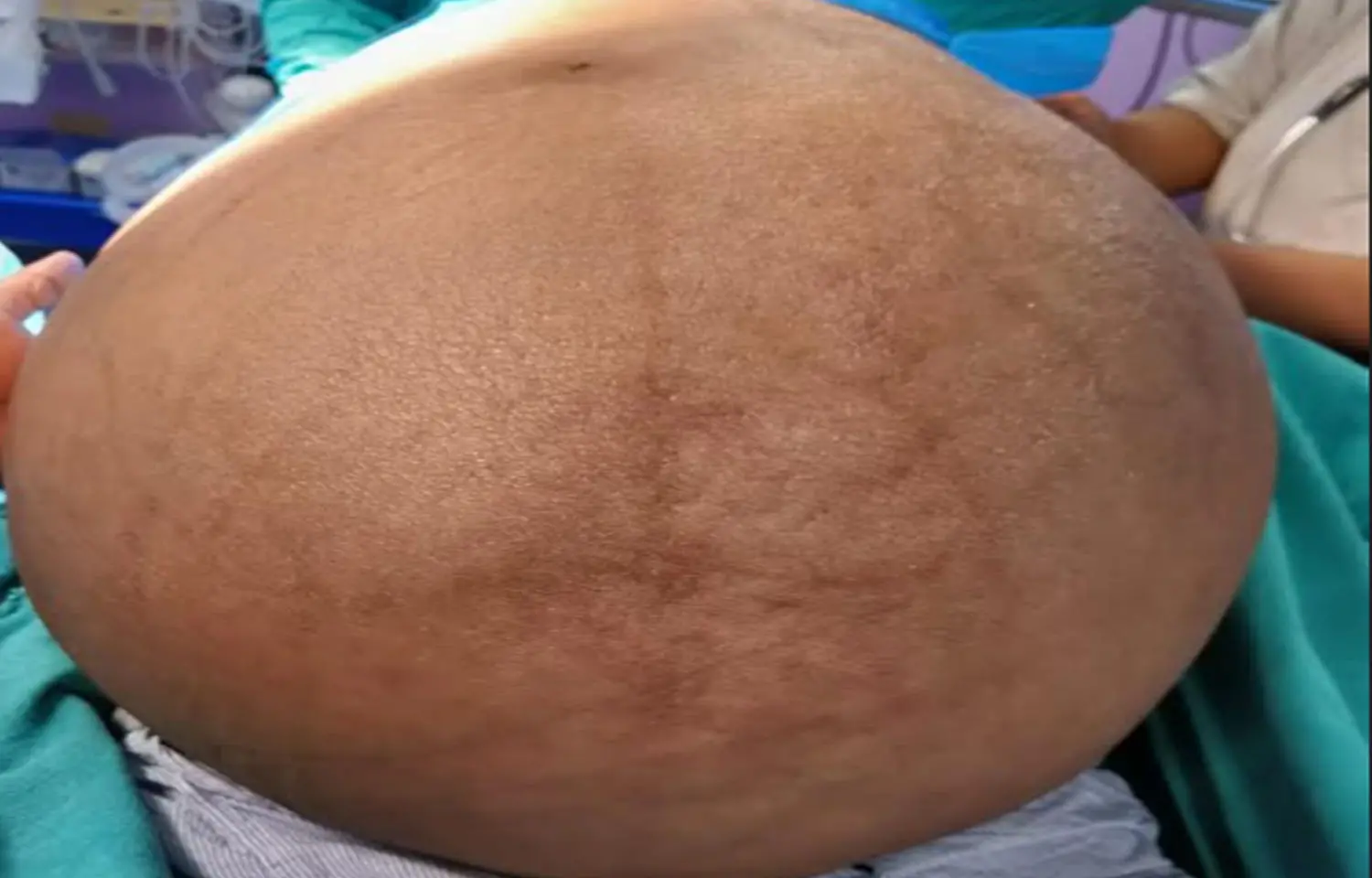
A 54-year-old woman (gravida 0, para 0) was transferred to our department with an extensively distended abdominal wall and leg pain. Regular menstruation started at age 14, and she experienced menopause at age 48. She had no history of regular hospitalizations. Over the past few years, she had noticed a gradual progression of abdominal bloating, but she had not decided to go to the hospital. Finally, when it became difficult for her to walk by herself, she went to a nearby hospital and was transferred to our department. Her vital signs were stable; however, her abdomen was markedly distended from the cardiac fossa to the lower abdomen, making it difficult for her to stand by herself. Marked pitting edema was found in both legs.
Contrast-enhanced computed tomography showed that the tumor occupied the whole abdominal cavity (38 cm × 40 cm × 48 cm), and both kidneys were being pressed significantly dorsally. Most of the tumor was uniform, and its density was like that of subcutaneous fat. There were no hypervascular lesions, and the right ovarian artery and vein flowed into the tumor. No obvious venous thrombosis was detected; however, pleural effusion was detected in the right thoracic region. The tumor was too large to obtain useful information from magnetic resonance imaging.
Blood test results showed that the CA125 value was slightly elevated, and there was a marked increase of estradiol and a marked suppression of luteinizing hormone and follicle stimulating hormone levels, which indicated a benign ovarian solid fibroma or thecoma with Meigs syndrome.
Authors planned to surgically remove the right adnexa, but because of concerns about potentially severe complications, they organized a multidisciplinary team of general surgeons, anesthetists, radiation oncologists, and plastic surgeons to plan the treatment course.
During the laparotomy, the patient was placed in the left lateral decubitus position to maintain hemodynamic stability and because the tumor was assumed to be of right ovarian origin. With the help of general surgeons, team confirmed that there were no adhesions between the tumor and the abdominal wall, and the surface of the tumor was smooth. They confirmed that the right ovarian artery and vein truly flowed into the tumor. Her uterine and left adnexa were intact. They cut both vessels, the right fallopian tube, and the ovarian intrinsic ligament and successfully removed the right adnexa. The tumor weighed 36 kg. Because the subcutaneous fascia and skin were markedly stretched by the tumor, a plastic surgeon trimmed the excess fascia and skin and reformed the umbilicus. During the operation, the patient's vital signs were fairly stable. The amount of intraoperative blood loss was 420 mL, and the operation time was 4 hours and 17 minutes.
The patient was then extubated and moved to the intensive care unit for recovery. There were no signs of major complications, and she was moved to the general ward on the 1st postoperative day. A chest radiograph on the fourth postoperative day showed a marked decrease in the right pleural effusion. The postoperative course was generally favorable, and the patient was discharged on the 7th postoperative day.
A pathological examination showed that the tumor was macroscopically nearly white, but there were no obvious necrotic lesions. Microscopically, the tumor was composed of thin spindle cells in a whorled arrangement, but nuclear atypia and mitosis were not observed, and the fibroma diagnosis was confirmed.
On the 29th postoperative day, the patient visited the outpatient, and the wound was observed to be healing well. A blood test performed 7 months after the surgery confirmed that her hormonal status had returned to the menopausal status, and she did not show any complaints.
Up to 25% of ExGOvTs could be malignant or borderline malignant. In addition, their age and tumor size were found not to be related to the frequency of malignancy or borderline malignancy. One should employ several diagnostic approaches, including imaging and physical examinations, to assess the malignancy potential of each case. In the present case, although some tumor markers were moderately elevated, contrast enhanced computed tomography showed a relatively homogenous area within the tumor and no obvious lymph node enlargement or tumor dissemination lesions. Therefore, a benign tumor with Meigs syndrome was suspected.
One should take the high preoperative mortality and the high frequency of fatal postoperative complication into account when determining the treatment strategy for ExG-OvTs regardless of their malignant potential. When surgical interventions are planned, it is obviously critical to employ a multidisciplinary team, including anesthesiologists, cardiologists, and general and plastic surgeons. In the present case, the surgery was fortunately completed without severe complications. For cystic tumors, the tumor fluid can gradually be aspirated to prevent rapid hemodynamic changes; however, this is not possible for solid tumors. In the present case, one did not occur.
"In conclusion, to the best of our knowledge, we successfully resected the largest solid ovarian tumor that has ever been reported with help of a multidisciplinary team without any severe complications. Although many ExG-OvTs (about 75%) are pathologically benign, they are still strongly associated with a high mortality and fatal postoperative complications, and we have to take that into account when planning the treatment."
Source: Miyu Tanaka , Koji Yamanoi , Sachiko Kitamura; Hindawi Case Reports in Obstetrics and Gynecology Volume 2021
https://doi.org/10.1155/2021/1076855
MBBS, MD Obstetrics and Gynecology
Dr Nirali Kapoor has completed her MBBS from GMC Jamnagar and MD Obstetrics and Gynecology from AIIMS Rishikesh. She underwent training in trauma/emergency medicine non academic residency in AIIMS Delhi for an year after her MBBS. Post her MD, she has joined in a Multispeciality hospital in Amritsar. She is actively involved in cases concerning fetal medicine, infertility and minimal invasive procedures as well as research activities involved around the fields of interest.
Dr Kamal Kant Kohli-MBBS, DTCD- a chest specialist with more than 30 years of practice and a flair for writing clinical articles, Dr Kamal Kant Kohli joined Medical Dialogues as a Chief Editor of Medical News. Besides writing articles, as an editor, he proofreads and verifies all the medical content published on Medical Dialogues including those coming from journals, studies,medical conferences,guidelines etc. Email: drkohli@medicaldialogues.in. Contact no. 011-43720751


