- Home
- Medical news & Guidelines
- Anesthesiology
- Cardiology and CTVS
- Critical Care
- Dentistry
- Dermatology
- Diabetes and Endocrinology
- ENT
- Gastroenterology
- Medicine
- Nephrology
- Neurology
- Obstretics-Gynaecology
- Oncology
- Ophthalmology
- Orthopaedics
- Pediatrics-Neonatology
- Psychiatry
- Pulmonology
- Radiology
- Surgery
- Urology
- Laboratory Medicine
- Diet
- Nursing
- Paramedical
- Physiotherapy
- Health news
- Fact Check
- Bone Health Fact Check
- Brain Health Fact Check
- Cancer Related Fact Check
- Child Care Fact Check
- Dental and oral health fact check
- Diabetes and metabolic health fact check
- Diet and Nutrition Fact Check
- Eye and ENT Care Fact Check
- Fitness fact check
- Gut health fact check
- Heart health fact check
- Kidney health fact check
- Medical education fact check
- Men's health fact check
- Respiratory fact check
- Skin and hair care fact check
- Vaccine and Immunization fact check
- Women's health fact check
- AYUSH
- State News
- Andaman and Nicobar Islands
- Andhra Pradesh
- Arunachal Pradesh
- Assam
- Bihar
- Chandigarh
- Chattisgarh
- Dadra and Nagar Haveli
- Daman and Diu
- Delhi
- Goa
- Gujarat
- Haryana
- Himachal Pradesh
- Jammu & Kashmir
- Jharkhand
- Karnataka
- Kerala
- Ladakh
- Lakshadweep
- Madhya Pradesh
- Maharashtra
- Manipur
- Meghalaya
- Mizoram
- Nagaland
- Odisha
- Puducherry
- Punjab
- Rajasthan
- Sikkim
- Tamil Nadu
- Telangana
- Tripura
- Uttar Pradesh
- Uttrakhand
- West Bengal
- Medical Education
- Industry
Anatomical and Functional Outcomes in Delayed Onset versus Concurrent Retinal Detachment in Endophthalmitis

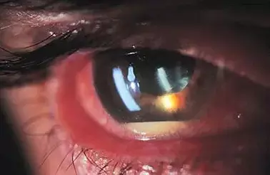
Endophthalmitis is a devastating condition characterized by a severe intraocular infection that is often associated with poor visual outcomes. Although retinal detachment (RD) has been described as an uncommon complication in endophthalmitis, an early diagnosis is imperative as it significantly affects the management strategies and visual outcomes in these patients. The postoperative functional and anatomical outcomes of RD in endophthalmitis are dependent on the etiology, virulence of the causative microorganisms, severity of intraocular inflammation, status of proliferative vitreoretinopathy (PVR), time of diagnosis, visual acuity (VA) at presentation, presence of an intraocular foreign body and timing of treatment. In the setting of endophthalmitis, RD may be either diagnosed as a part of initial presentation or noted on follow-up after the therapeutic vitrectomy procedure.
The aim of the study by Srinivasan et al was to determine the functional and anatomical outcomes of patients with endophthalmitis with concurrent or delayed onset RD, and to compare the preoperative, intraoperative and postoperative features in these two presentations of RD in patients with endophthalmitis.
This was a retrospective review of 121 eyes in 121 patients presenting with endophthalmitis and RD. Subjects were categorized into two groups: endophthalmitis with delayed onset RD (group 1, N=76) and endophthalmitis with concurrent RD (group 2, N=45).
Exogenous endophthalmitis was common in both groups 1 and 2 (86.84% and 84.44%, respectively). No significant differences were found between the groups in the type of RD, retinal breaks, number of quadrants involved or proliferative vitreoretinopathy grade. In the overall cohort, visual acuity improved post-surgery in one-third of the patients who were in the near or total blindness category at presentation. Authors found good anatomical success rates of an attached retina in both groups 1 and 2 (84.3% and 77.7%, P=0.376).
This study is a comparative analysis of patients with endophthalmitis diagnosed with either delayed onset or concurrent RD. Authors compared the demographic, microbiological and RD characteristics, and reported anatomical and functional success in both groups. The patients in group 1 (delayed onset RD) were slightly younger than the patients in group 2 (concurrent RD). The mean age at presentation was in the late 30s for group 1 and the late 40s for group 2. The etiology of endophthalmitis in both groups was found to be exogenous in >80% of the study patients. While cataract surgery and trauma was noted as the most common etiology for exogenous endophthalmitis in group 1, cataract surgery alone was the predominant etiology in group 2.
“Our study presents the outcomes in patients with endophthalmitis with concurrent or delayed onset RD. Although the presenting visual acuity and status of the retina showed significant variation at presentation, the anatomical and functional outcomes were almost the same. Hence, the visual acuity and final retinal status did not differ significantly based on whether the RD was concurrent or delayed onset in nature.”
Source: Srinivasan et al; Clinical Ophthalmology 2023:17 115–121
https://doi.org/10.2147/OPTH.S389474
Dr Ishan Kataria has done his MBBS from Medical College Bijapur and MS in Ophthalmology from Dr Vasant Rao Pawar Medical College, Nasik. Post completing MD, he pursuid Anterior Segment Fellowship from Sankara Eye Hospital and worked as a competent phaco and anterior segment consultant surgeon in a trust hospital in Bathinda for 2 years.He is currently pursuing Fellowship in Vitreo-Retina at Dr Sohan Singh Eye hospital Amritsar and is actively involved in various research activities under the guidance of the faculty.
Dr Kamal Kant Kohli-MBBS, DTCD- a chest specialist with more than 30 years of practice and a flair for writing clinical articles, Dr Kamal Kant Kohli joined Medical Dialogues as a Chief Editor of Medical News. Besides writing articles, as an editor, he proofreads and verifies all the medical content published on Medical Dialogues including those coming from journals, studies,medical conferences,guidelines etc. Email: drkohli@medicaldialogues.in. Contact no. 011-43720751


