- Home
- Medical news & Guidelines
- Anesthesiology
- Cardiology and CTVS
- Critical Care
- Dentistry
- Dermatology
- Diabetes and Endocrinology
- ENT
- Gastroenterology
- Medicine
- Nephrology
- Neurology
- Obstretics-Gynaecology
- Oncology
- Ophthalmology
- Orthopaedics
- Pediatrics-Neonatology
- Psychiatry
- Pulmonology
- Radiology
- Surgery
- Urology
- Laboratory Medicine
- Diet
- Nursing
- Paramedical
- Physiotherapy
- Health news
- Fact Check
- Bone Health Fact Check
- Brain Health Fact Check
- Cancer Related Fact Check
- Child Care Fact Check
- Dental and oral health fact check
- Diabetes and metabolic health fact check
- Diet and Nutrition Fact Check
- Eye and ENT Care Fact Check
- Fitness fact check
- Gut health fact check
- Heart health fact check
- Kidney health fact check
- Medical education fact check
- Men's health fact check
- Respiratory fact check
- Skin and hair care fact check
- Vaccine and Immunization fact check
- Women's health fact check
- AYUSH
- State News
- Andaman and Nicobar Islands
- Andhra Pradesh
- Arunachal Pradesh
- Assam
- Bihar
- Chandigarh
- Chattisgarh
- Dadra and Nagar Haveli
- Daman and Diu
- Delhi
- Goa
- Gujarat
- Haryana
- Himachal Pradesh
- Jammu & Kashmir
- Jharkhand
- Karnataka
- Kerala
- Ladakh
- Lakshadweep
- Madhya Pradesh
- Maharashtra
- Manipur
- Meghalaya
- Mizoram
- Nagaland
- Odisha
- Puducherry
- Punjab
- Rajasthan
- Sikkim
- Tamil Nadu
- Telangana
- Tripura
- Uttar Pradesh
- Uttrakhand
- West Bengal
- Medical Education
- Industry
AREDS 5-step Simplified Severity Scale for Macular Degeneration Classification- a Novel Design

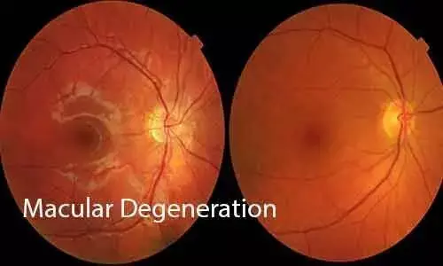
Age-related macular degeneration (AMD) is responsible for approximately 9% of global blindness and is the leading cause of visual loss in developed countries. AMD is a progressive, stepwise disease and is classified, based on clinical examination or color fundus photography, into early, intermediate, and late stages. The hallmarks of intermediate disease are the presence of large drusen or pigmentary abnormalities at the macula. There are two forms of late AMD: 1) neovascular AMD and 2) atrophic AMD, with geographic atrophy (GA).
The Age-Related Eye Disease Study (AREDS), sponsored by the National Eye Institute (National Institutes of Health), was a randomized clinical trial to assess the efficacy of oral supplementation with antioxidant vitamins and minerals on the clinical course of AMD and age-related cataract. Longitudinal analysis of this study cohort led to the development of the patient-based AREDS Simplified Severity Scale for AMD. It combines risk factors from both eyes to generate an overall score for the individual, based on the presence of one or more large drusen (diameter >125 mm) or pigmentary abnormalities at the macula of each eye. The 5-step Simplified Severity Scale can be used by clinicians to predict an individual’s 5-year risk of developing late AMD and thus determine which patient would benefit from anti-oxidant supplements to reduce the risk of them developing neovascular AMD. The rising disease prevalence will place a significant burden on existing eye care services, and as a result there is increasing interest in the use of automated diagnostics and tele-ophthalmology services to identify at-risk individuals and facilitate both the early diagnosis of sight-threatening disease and instigation of early intervention strategies to reduce the risk of disease progression.
This approach is of interest because if this scale could be generated automatically from retinal images it would enable the development of tools that could enable mass screening for AMD in primary care, without recourse to specialist review. In the current paper, we hypothesized that it should be possible to build CNNs that would more accurately detect those elements critical to the AREDS 5-step Simplified Severity Scale, namely advanced AMD (aAMD), drusen size and retinal pigmentary abnormalities, if higher quality images were used. We also investigated the effect of image size, network architecture and hyperspace optimization on the overall performance of the neural network. The accuracy of the resulting optimized neural network to predict the final 5-step severity scale score generated was then assessed against the ground truth using traditional metrics, ROC analysis, sensitivity and specificity.
This was a Retrospective cohort study. Three individual CNNs were trained to accurately detect 1) advanced AMD, 2) drusen size and 3) the presence or otherwise of pigmentary abnormalities, from macular centered retinal images were developed. The CNNs were then arranged in a “cascading” architecture to calculate the Age-related Eye Disease Study (AREDS) Simplified 5-level risk Severity score (Risk Score 0 – Risk Score 4), for test images. The process was repeated creating a simplified binary “low risk” (Scores 0–2) and “high risk” (Risk Score 3– 4) classification.
There were a total of 188,006 images, of which 118,254 images were deemed gradable, representing 4591 patients, from the AREDS1 dataset. The gradable images were split into 50%/25%/25% ratios for training, validation and test purposes.
When assessed against the 5-step Simplified Severity Scale, the results generated by the ensemble of CNN’s achieved an accuracy of 80.43%. When assessed against a simplified binary (Low Risk/High Risk) classification, an accuracy of 98.08%, sensitivity of ≥85% and specificity of ≥99% was achieved.
The AREDS 5-step Simplified Severity Scale was developed to provide clinically useful risk categories for the development of advanced AMD in persons with earlier stages of AMD. It results in a scoring system that assigns 1 point for the presence of one or more large (≥125 µm) drusen, 1 point for the presence of any retinal pigment abnormalities in an eye, and 1 point for bilateral medium drusen if there are no large drusen in either eye. Risk factors are summed across both eyes, forming a 5-step scale (steps 0–4) for which the 5-year risk of developing advanced AMD in at least 1 eye is derived.
Authors used a novel CNN approach to automate the classification of the images in the AREDS dataset with a view to identifying the three principal components of the Simplified Severity score: advanced AMD, large drusen and pigmentary abnormalities with the aim of then being able to automate the risk of an individual’s disease progressing using the AREDS 5-step Simplified Severity Scale Scores. At the patient level, the classifier achieved a 5-class accuracy of 78.49% and 80.43%, and a quadratic kappa of 0.854 and 0.870 for the 600*600 images and 800*800 images, respectively.
If the 5-class-AREDS Simplified scale is further reduced to a binary low risk/high risk outcome, the accuracy of our combined neural networks was 98% with a sensitivity of ≥85% and a specificity ≥99% for both the 600*600 and the 800*800 images. The improvement in neural network performance with image size is in keeping with published data. Whilst increasing image size leads to significant efficacy gains between 200*200 and 500*500 pixels sizes, these gains quickly plateau when larger sized images (800*800 to 2000*2000) are used.
Authors have created individual neural networks, trained on macular centered images from the AREDS 1 dataset, that are capable of accurately grading discrete clinical features of AMD from macular centered fundal images. When arranged in a cascading ensemble, the grades issued by these individual networks allowed us to accurately calculate the AREDS 5-step Simplified Severity Scale score for AMD for any given individual. If the results presented were replicated, then the ensemble of neural networks they have developed could be used as a screening tool that has the potential to significantly improve health outcomes by identifying asymptomatic individuals who would benefit from AREDS2 macular supplements.
Source: Xie et al; Clinical Ophthalmology 2023:17
https://doi.org/10.2147/OPTH.S396537
Dr Ishan Kataria has done his MBBS from Medical College Bijapur and MS in Ophthalmology from Dr Vasant Rao Pawar Medical College, Nasik. Post completing MD, he pursuid Anterior Segment Fellowship from Sankara Eye Hospital and worked as a competent phaco and anterior segment consultant surgeon in a trust hospital in Bathinda for 2 years.He is currently pursuing Fellowship in Vitreo-Retina at Dr Sohan Singh Eye hospital Amritsar and is actively involved in various research activities under the guidance of the faculty.
Dr Kamal Kant Kohli-MBBS, DTCD- a chest specialist with more than 30 years of practice and a flair for writing clinical articles, Dr Kamal Kant Kohli joined Medical Dialogues as a Chief Editor of Medical News. Besides writing articles, as an editor, he proofreads and verifies all the medical content published on Medical Dialogues including those coming from journals, studies,medical conferences,guidelines etc. Email: drkohli@medicaldialogues.in. Contact no. 011-43720751


