
Autologous Blood Clotting Seals Unanticipated Posterior Scleral Perforation During Vitrectomy: Case Report
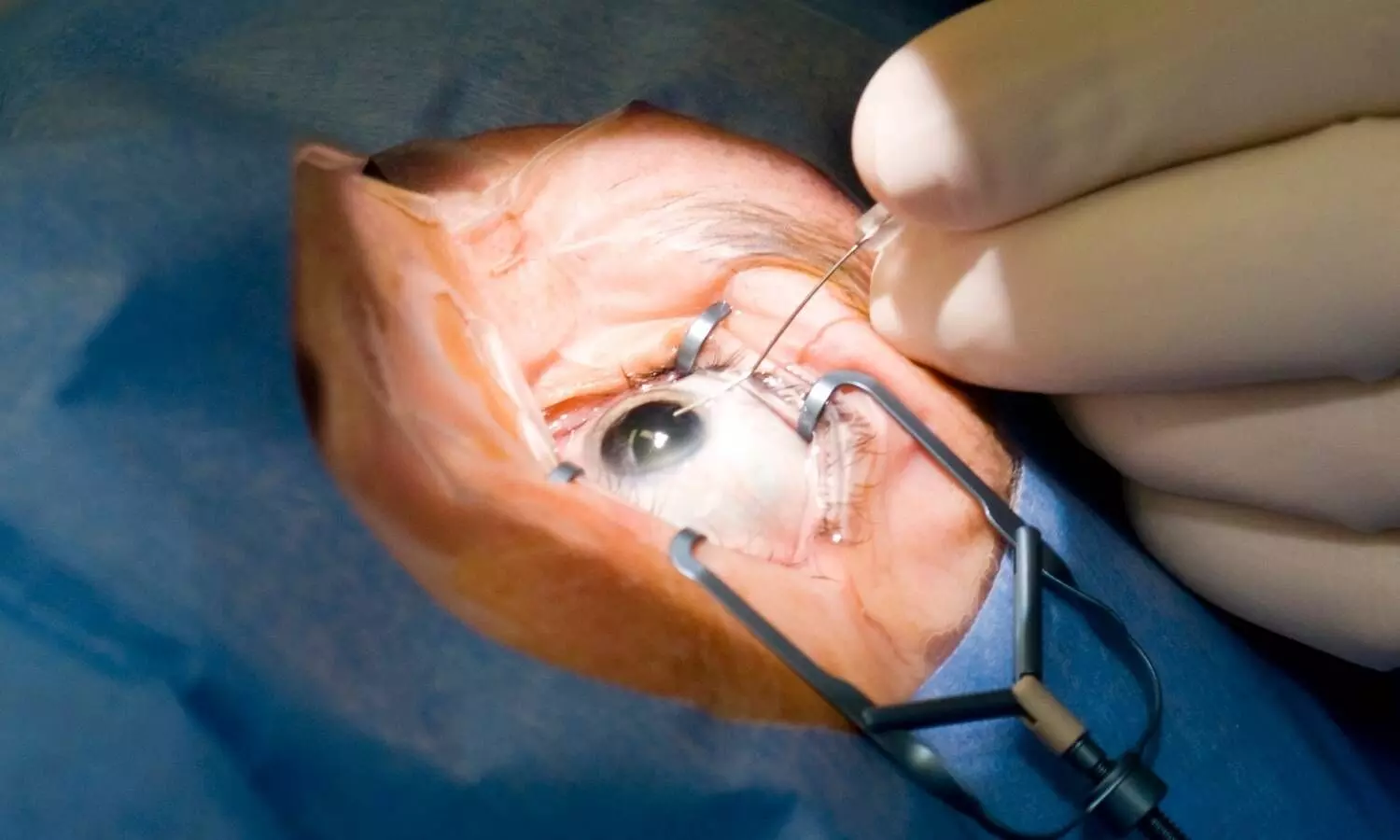


A recent case report introduces a simple rescue technique that uses an autologous blood clot to seal unexpected posterior scleral perforations during vitreoretinal surgery.
The findings were published in May 2026 in the Indian Journal of Ophthalmology by Dr. Günhal Şatırtav and colleagues from Necmettin Erbakan University School of Medicine in Türkiye.
A 34-year-old male presented with penetrating ocular trauma from a gunshot injury, featuring a radiopaque intraocular foreign body deeply embedded in the posterior scleral wall. The patient underwent primary corneal reconstruction. Following a seven-day waiting period, surgeons performed a three-port 25-gauge pars plana vitrectomy. After core vitrectomy and posterior hyaloid detachment, the foreign body was extracted from its intrascleral bed. During fluid-air exchange, surgeons observed massive subconjunctival and sub-Tenon air accumulation, confirming an active air leak from the posterior scleral defect.
To address the unanticipated defect without specialized graft materials, surgeons executed a rapid autologous blood-assisted closure:
- Venipuncture and Application: Approximately 0.2 to 0.3 mL of autologous venous blood was drawn from the patient's antecubital vein and applied over the posterior defect using a cannula under air.
- Clot Positioning and Stabilization: Approximately 0.1 mL of blood was clotted directly over the perforation. Clotted blood pooling near the optic disc was repositioned over the defect, and intraocular fluid was aspirated to establish a stable biological seal.
- Laser Photocoagulation: Endolaser photocoagulation was delivered directly onto the blood clot to stabilize the fibrin matrix and onto the surrounding retina.
- Gas Tamponade: Perfluoropropane (C₃F₈) gas was injected as an intraocular tamponade to maintain pressure against the clot during initial healing.
Mechanistic Insights
Posterior scleral dehiscence after foreign body extraction poses severe surgical risks, including intraocular fluid escape, orbital air accumulation, hypotony, and air embolism. Traditional management strategies require demanding ab interno suturing or donor materials like amniotic membranes, temporal fascia, or donor sclera. In contrast, autologous blood provides an immediate biological glue rich in fibrinogen. This approach eliminates graft rejection and material acquisition delays while reducing technical complexity. Postoperatively, ocular structural integrity was successfully restored alongside improved visual outcomes.
Implications for Ophthalmic Surgeons
Ophthalmic surgeons should consider autologous blood application as a reliable rescue strategy for unexpected posterior scleral perforations encountered during vitrectomy. This technique equips retina specialists with an accessible, cost-effective tool to salvage posterior pole integrity when conventional tissue grafts are unavailable.
Reference
Şatırtav, G., Kerimoğlu, H., & Adam, M. (2026). Autologous blood-assisted closure of an unanticipated posterior scleral perforation: A novel rescue technique. Indian Journal of Ophthalmology, 74(Suppl 3), S471–S472.