- Home
- Medical news & Guidelines
- Anesthesiology
- Cardiology and CTVS
- Critical Care
- Dentistry
- Dermatology
- Diabetes and Endocrinology
- ENT
- Gastroenterology
- Medicine
- Nephrology
- Neurology
- Obstretics-Gynaecology
- Oncology
- Ophthalmology
- Orthopaedics
- Pediatrics-Neonatology
- Psychiatry
- Pulmonology
- Radiology
- Surgery
- Urology
- Laboratory Medicine
- Diet
- Nursing
- Paramedical
- Physiotherapy
- Health news
- Fact Check
- Bone Health Fact Check
- Brain Health Fact Check
- Cancer Related Fact Check
- Child Care Fact Check
- Dental and oral health fact check
- Diabetes and metabolic health fact check
- Diet and Nutrition Fact Check
- Eye and ENT Care Fact Check
- Fitness fact check
- Gut health fact check
- Heart health fact check
- Kidney health fact check
- Medical education fact check
- Men's health fact check
- Respiratory fact check
- Skin and hair care fact check
- Vaccine and Immunization fact check
- Women's health fact check
- AYUSH
- State News
- Andaman and Nicobar Islands
- Andhra Pradesh
- Arunachal Pradesh
- Assam
- Bihar
- Chandigarh
- Chattisgarh
- Dadra and Nagar Haveli
- Daman and Diu
- Delhi
- Goa
- Gujarat
- Haryana
- Himachal Pradesh
- Jammu & Kashmir
- Jharkhand
- Karnataka
- Kerala
- Ladakh
- Lakshadweep
- Madhya Pradesh
- Maharashtra
- Manipur
- Meghalaya
- Mizoram
- Nagaland
- Odisha
- Puducherry
- Punjab
- Rajasthan
- Sikkim
- Tamil Nadu
- Telangana
- Tripura
- Uttar Pradesh
- Uttrakhand
- West Bengal
- Medical Education
- Industry
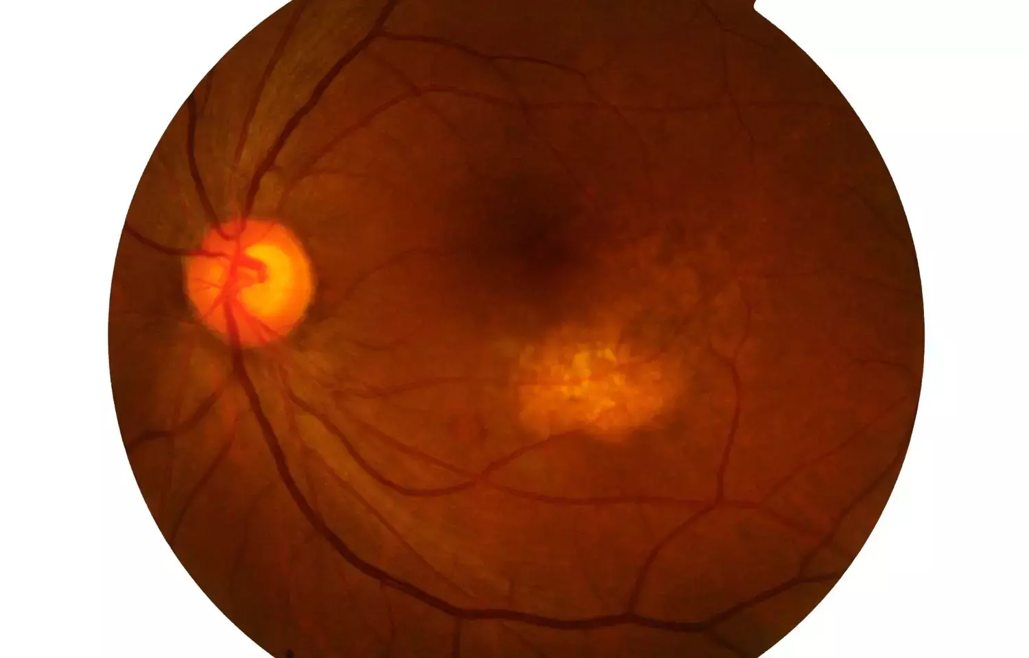
Color FAF imaging - A novel diagnostic Biomarker for Posterior Uveitis Lesions

Spectrally resolved fundus autofluorescence can be used as a novel diagnostic biomarker for Posterior Uveitis lesions states a study that was published in the journal Scientific Reports.
It is quite a challenge to clinically discriminate posterior uveitis lesions as most of the posterior uveitis lesions are not clearly understood. Spectrally resolved Fundus autofluorescence (Color-FAF) is a novel, non-invasive method that has been used in detecting various retinal lesions. Hence, researchers from the University Hospital Bonn, Department of Ophthalmology, Germany, conducted a study to evaluate Color-FAF imaging as a diagnostic imaging biomarker in different posterior uveitis entities. An exploratory, retrospective, cross-sectional study was conducted on patients with posterior uveitis. Green (GEFC) and red emission fluorescent components (REFC) of retinal and choroidal lesions in posterior uveitis were investigated to facilitate discrimination of the different entities.
Optical coherence tomography, Color fundus photography, and spectrally resolved fundus autofluorescence (Color-FAF) was used to image the eyes. Retinal/choroidal lesions' intensities of GEFC (500–560 nm) and REFC (560–700 nm) were determined, and intensity-normalized Color-FAF images were compared for birdshot chorioretinopathy, ocular sarcoidosis, acute posterior multifocal placoid pigment epitheliopathy (APMPPE), and punctate inner choroidopathy (PIC). Possible confounders were revealed using Multivariable regression analyses.
Key Findings:
- 76 eyes of 45 patients were included with a total of 845 lesions.
- Mean GEFC/REFC ratios were 0.82 ± 0.10, 0.92 ± 0.11, 0.86 ± 0.10, and 1.09 ± 0.19 for birdshot chorioretinopathy, sarcoidosis, APMPPE, and PIC lesions, respectively, and were significantly different in repeated measures ANOVA (p < 0.0001).
- Non-pigmented retinal/choroidal lesions, macular neovascularizations, and fundus areas of choroidal thinning featured predominantly GEFC, and pigmented retinal lesions predominantly REFC.
- Color-FAF imaging revealed the involvement of both, short- and long-wavelength emission fluorophores in posterior uveitis.
- The GEFC/REFC ratio of retinal and choroidal lesions was significantly different between distinct subgroups.
Thus, the researchers concluded that for differentiation of posterior uveitis lesions spectrally resolved fundus autofluorescence (Color-FAF) can be remarkably used as a new non-invasive diagnostic tool.
For further reading: https://doi.org/10.1038/s41598-022-18048-4
Wintergerst, M.W.M., Merten, N.R., Berger, M. et al. Spectrally resolved autofluorescence imaging in posterior uveitis. Sci Rep 12, 14337 (2022).
BDS, MDS
Dr.Niharika Harsha B (BDS,MDS) completed her BDS from Govt Dental College, Hyderabad and MDS from Dr.NTR University of health sciences(Now Kaloji Rao University). She has 4 years of private dental practice and worked for 2 years as Consultant Oral Radiologist at a Dental Imaging Centre in Hyderabad. She worked as Research Assistant and scientific writer in the development of Oral Anti cancer screening device with her seniors. She has a deep intriguing wish in writing highly engaging, captivating and informative medical content for a wider audience. She can be contacted at editorial@medicaldialogues.in.
Dr Kamal Kant Kohli-MBBS, DTCD- a chest specialist with more than 30 years of practice and a flair for writing clinical articles, Dr Kamal Kant Kohli joined Medical Dialogues as a Chief Editor of Medical News. Besides writing articles, as an editor, he proofreads and verifies all the medical content published on Medical Dialogues including those coming from journals, studies,medical conferences,guidelines etc. Email: drkohli@medicaldialogues.in. Contact no. 011-43720751


