- Home
- Medical news & Guidelines
- Anesthesiology
- Cardiology and CTVS
- Critical Care
- Dentistry
- Dermatology
- Diabetes and Endocrinology
- ENT
- Gastroenterology
- Medicine
- Nephrology
- Neurology
- Obstretics-Gynaecology
- Oncology
- Ophthalmology
- Orthopaedics
- Pediatrics-Neonatology
- Psychiatry
- Pulmonology
- Radiology
- Surgery
- Urology
- Laboratory Medicine
- Diet
- Nursing
- Paramedical
- Physiotherapy
- Health news
- Fact Check
- Bone Health Fact Check
- Brain Health Fact Check
- Cancer Related Fact Check
- Child Care Fact Check
- Dental and oral health fact check
- Diabetes and metabolic health fact check
- Diet and Nutrition Fact Check
- Eye and ENT Care Fact Check
- Fitness fact check
- Gut health fact check
- Heart health fact check
- Kidney health fact check
- Medical education fact check
- Men's health fact check
- Respiratory fact check
- Skin and hair care fact check
- Vaccine and Immunization fact check
- Women's health fact check
- AYUSH
- State News
- Andaman and Nicobar Islands
- Andhra Pradesh
- Arunachal Pradesh
- Assam
- Bihar
- Chandigarh
- Chattisgarh
- Dadra and Nagar Haveli
- Daman and Diu
- Delhi
- Goa
- Gujarat
- Haryana
- Himachal Pradesh
- Jammu & Kashmir
- Jharkhand
- Karnataka
- Kerala
- Ladakh
- Lakshadweep
- Madhya Pradesh
- Maharashtra
- Manipur
- Meghalaya
- Mizoram
- Nagaland
- Odisha
- Puducherry
- Punjab
- Rajasthan
- Sikkim
- Tamil Nadu
- Telangana
- Tripura
- Uttar Pradesh
- Uttrakhand
- West Bengal
- Medical Education
- Industry
Pseudoexfoliation Syndrome May Predict Glaucoma via Ganglion Cell Layer Thinning: Study

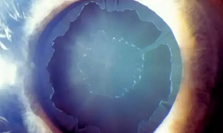
Portugal: Researchers have found in a cross-sectional study from Portugal that pseudoexfoliation (PEX) syndrome is linked to early thinning of the ganglion cell layer (GCL) and inner plexiform layer (IPL), suggesting these changes may serve as early predictors of glaucoma development.
The study, conducted by Dr. Ana Faria Pereira and colleagues from the Department of Ophthalmology, Unidade Local de Saúde de São João, Porto, and published in Scientific Reports, sheds light on the structural retinal alterations in PEX patients, even before signs of glaucoma appear.
PEX syndrome is characterized by the accumulation of fibrillar material within ocular and systemic tissues, primarily involving the eye's anterior segment. While it is already established as a major risk factor for open-angle glaucoma, its broader effects on the retina remain underexplored.
To address this, the researchers examined macular, choroidal, and circumpapillary retinal nerve fiber layer (cRNFL) parameters using spectral-domain optical coherence tomography (SD-OCT). The study included 60 eyes—38 affected by PEX and 22 from healthy controls. All participants had intraocular pressures (IOP) below 19 mmHg and showed no clinical signs of glaucomatous damage.
The study led to the following findings:
- PEX patients exhibited a significant reduction in nasal-inferior circumpapillary retinal nerve fiber layer (cRNFL) thickness.
- The ganglion cell layer (GCL) in the superior, inferior, nasal-inferior, nasal-superior, and total macular regions was notably thinner in the PEX group.
- Corresponding inner plexiform layer (IPL) areas in these regions also showed significant thinning in PEX patients.
- The Bruch’s membrane opening-minimum rim width (BMO-MRW) was slightly thinner across all sectors in the PEX group, though this difference was not statistically significant.
- Temporal choroidal thickness was significantly higher in PEX eyes, contrary to typical patterns seen in degenerative conditions.
- These findings indicate that PEX may contribute to early retinal neurodegeneration, even without elevated intraocular pressure or evident glaucomatous damage.
- The nasal regions of the GCL and IPL may serve as valuable areas for early detection of preclinical glaucoma or other neurodegenerative disorders.
The study does note several limitations, including potential variability in IOP and the inclusion of both eyes from some participants, which may influence data consistency. Additionally, despite statistically significant thinning in retinal layers, values remained within normal ranges, emphasizing the need for longitudinal assessment to determine progression.
The authors advocate for larger-scale, prospective studies that incorporate brain imaging and dementia evaluations to better understand how retinal changes in PEX may parallel or predict broader neurological conditions. These findings underscore the potential of SD-OCT imaging in detecting early neuroretinal damage, which may enhance early glaucoma diagnosis and even help screen for systemic neurodegenerative diseases.
Reference:
Faria Pereira, A., Mota Moreira, P., & Estrela Silva, S. (2025). Retinal neurodegeneration, neuroretinal rim analysis and choroid thickness in pseudoexfoliation syndrome with spectral domain optical coherence tomography. Scientific Reports, 15(1), 1-9. https://doi.org/10.1038/s41598-025-95082-y
Dr Kamal Kant Kohli-MBBS, DTCD- a chest specialist with more than 30 years of practice and a flair for writing clinical articles, Dr Kamal Kant Kohli joined Medical Dialogues as a Chief Editor of Medical News. Besides writing articles, as an editor, he proofreads and verifies all the medical content published on Medical Dialogues including those coming from journals, studies,medical conferences,guidelines etc. Email: drkohli@medicaldialogues.in. Contact no. 011-43720751


