- Home
- Medical news & Guidelines
- Anesthesiology
- Cardiology and CTVS
- Critical Care
- Dentistry
- Dermatology
- Diabetes and Endocrinology
- ENT
- Gastroenterology
- Medicine
- Nephrology
- Neurology
- Obstretics-Gynaecology
- Oncology
- Ophthalmology
- Orthopaedics
- Pediatrics-Neonatology
- Psychiatry
- Pulmonology
- Radiology
- Surgery
- Urology
- Laboratory Medicine
- Diet
- Nursing
- Paramedical
- Physiotherapy
- Health news
- Fact Check
- Bone Health Fact Check
- Brain Health Fact Check
- Cancer Related Fact Check
- Child Care Fact Check
- Dental and oral health fact check
- Diabetes and metabolic health fact check
- Diet and Nutrition Fact Check
- Eye and ENT Care Fact Check
- Fitness fact check
- Gut health fact check
- Heart health fact check
- Kidney health fact check
- Medical education fact check
- Men's health fact check
- Respiratory fact check
- Skin and hair care fact check
- Vaccine and Immunization fact check
- Women's health fact check
- AYUSH
- State News
- Andaman and Nicobar Islands
- Andhra Pradesh
- Arunachal Pradesh
- Assam
- Bihar
- Chandigarh
- Chattisgarh
- Dadra and Nagar Haveli
- Daman and Diu
- Delhi
- Goa
- Gujarat
- Haryana
- Himachal Pradesh
- Jammu & Kashmir
- Jharkhand
- Karnataka
- Kerala
- Ladakh
- Lakshadweep
- Madhya Pradesh
- Maharashtra
- Manipur
- Meghalaya
- Mizoram
- Nagaland
- Odisha
- Puducherry
- Punjab
- Rajasthan
- Sikkim
- Tamil Nadu
- Telangana
- Tripura
- Uttar Pradesh
- Uttrakhand
- West Bengal
- Medical Education
- Industry
"Two-Flaps" Technique- safe, reproducible, and consistent means for Descemet Stripping: Study

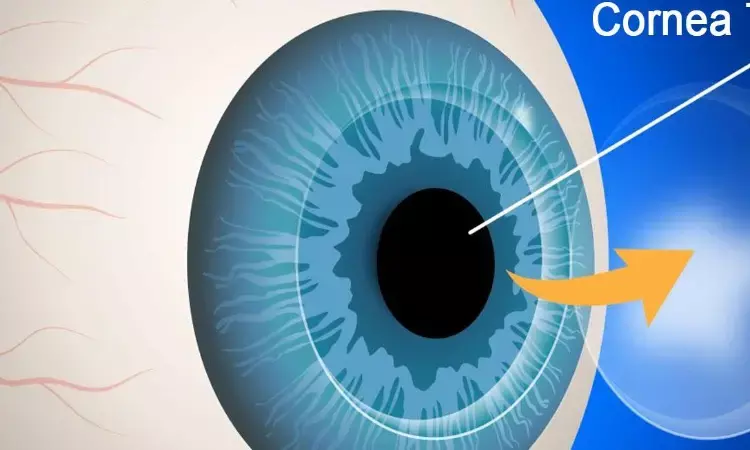
Fuchs endothelial dystrophy (FED) remains the most common indication for corneal transplantation in the United States. Surgical management of FED has undergone multiple iterations over the recent decades from initially performing penetrating keratoplasties to its current state of endothelial keratoplasty, which includes Descemet stripping automated endothelial keratoplasty and Descemet membrane endothelial keratoplasty. The evolution of surgical management of FED has now continued to progress to Descemet stripping only (DSO) in appropriate cases. DSO has gained popularity after observations of endothelial "rejuvenation" in FED despite complete Descemet membrane endothelial keratoplasty graft detachment. The proposed mechanism for this phenomenon was thought to be either migration and regeneration of the host endothelium or transfer from the donor endothelium free-floating graft.
There are a number of critical steps in the DSO surgical technique that must be implemented to optimize surgical success. These include optimal Descemet stripping centration, size of descemetorhexis, and achieving a smooth curvilinear descemetorhexis edge. Depending on the techniques, current success rates for DSO range between 63% and 100%. Currently, there is no step-by-step description of a safe and reproducible technique for the removal of Descemet membrane in the literature during DSO. Authors describe their current technique, the "two-flaps" technique, which they felt allowed for accurate, consistent, and minimally disruptive peeling of Descemet membrane.
An ink-marked caliper set at 4.0 mm was applied to the dry surface of the cornea with 8 points applied centrally over the nondilated pupil. The "two-flaps" technique uses the Gorovoy DSO forceps. The technique takes advantage of the flat and smooth surface of the forceps to create the desired 4-mm Descemet stripping with minimal stromal trauma along with a continuous curvilinear descemetorhexis, minimizing the risk of postoperative stromal scarring and extension of the rhexis beyond 4 mm. Holding the Gorovoy forceps closed and taking advantage of its wide and flat surface area, a small nick in Descemet membrane was made using the friction of the metal against Descemet membrane. This nick creation was first performed in an anticlockwise fashion and once formed, the Gorovoy forceps was then opened and grasped to encourage further guided peeling along the preplaced marking points, creating a 2 clock hours flap. With the opening of Descemet already created, the Gorovoy forceps are then used to peel in a clockwise manner for another 2 clock hours with the newly formed 4 clock hour Descemet crescent-shaped flap opening, Descemet was grasped in the middle of this opening and peeled toward the center of the 4-mm circumference up to the halfway point. The Gorovoy forceps was then used to perform a continuous curvilinear descemetorhexis, keeping within the 8-point marker. Once this was completed, the Descemet tissue was removed from the eye through the main wound. A well-centered descemetorhexis on visual axis was performed.
This technique has been used successfully in 11 cases performed by 1 surgeon or directly supervised by him. All cases achieved the desired 4-mm circumference without any residual tags or visually significant stromal scarring, with successful clearing of the central cornea and endothelial cells repopulating the central stripped area.
Peeling of Descemet membrane rather than scoring with inadvertent excessive pressure into the deeper stroma is important for surgical success. It has been observed that avpostoperative deep stromal scar usually corresponds to the location of where the scoring has taken place. The theory, therefore, is that the surgical trauma induced by the reverse Sinskey hook to score the endothelium/Descemet membrane complex initiates an inflammatory proscarring response, leading to stromal keratocyte activation, proliferation, fibrosis, and deep Stromal scarring.
The technique described reduces the complications associated with the use of a reverse Sinskey Hook. Using the smooth, nonsharp surface of the Gorovoy forceps for creating the first nick in Descemet membrane which then extends, by peeling only, to 2 flaps of 2 clock hours for each side reduces trauma to the underlying stroma and thereby reduces the risks of stromal scarring and stromal trench formation. This technique described provides a consistent, reproducible, and relatively trauma-free peeling of Descemet membrane and associated endothelial cells/guttae to optimize the success of DSO.
This "two-flaps" technique provides a broad edge of Descemet membrane to grip with initial 4 clock hours opening in Descemet membrane, which enhances peeling of a single continuous curvilinear sheet in a more controlled and accurate manner to the desired diameter size. Akin to a capsulorhexis, the surgeon has better control to manipulate the Descemet tear at the appropriate angle to ensure no tags or Descemet membrane remnants are left behind.
In conclusions, the "two-flap" technique described provides a safe, reproducible, and consistent means of performing a 4-mm descemetorhexis, which limits the trauma and possible subsequent scarring thus optimizing the success of DSO.
Source: Cohen, Eyal; Din, Nizar; Mimouni, Michael MD; Trinh, Tanya MD et al;
Cornea: September 2021 - Volume 40 - Issue 9 - p 1211-1214
doi: 10.1097/ICO.0000000000002744
Dr Ishan Kataria has done his MBBS from Medical College Bijapur and MS in Ophthalmology from Dr Vasant Rao Pawar Medical College, Nasik. Post completing MD, he pursuid Anterior Segment Fellowship from Sankara Eye Hospital and worked as a competent phaco and anterior segment consultant surgeon in a trust hospital in Bathinda for 2 years.He is currently pursuing Fellowship in Vitreo-Retina at Dr Sohan Singh Eye hospital Amritsar and is actively involved in various research activities under the guidance of the faculty.
Dr Kamal Kant Kohli-MBBS, DTCD- a chest specialist with more than 30 years of practice and a flair for writing clinical articles, Dr Kamal Kant Kohli joined Medical Dialogues as a Chief Editor of Medical News. Besides writing articles, as an editor, he proofreads and verifies all the medical content published on Medical Dialogues including those coming from journals, studies,medical conferences,guidelines etc. Email: drkohli@medicaldialogues.in. Contact no. 011-43720751


