- Home
- Medical news & Guidelines
- Anesthesiology
- Cardiology and CTVS
- Critical Care
- Dentistry
- Dermatology
- Diabetes and Endocrinology
- ENT
- Gastroenterology
- Medicine
- Nephrology
- Neurology
- Obstretics-Gynaecology
- Oncology
- Ophthalmology
- Orthopaedics
- Pediatrics-Neonatology
- Psychiatry
- Pulmonology
- Radiology
- Surgery
- Urology
- Laboratory Medicine
- Diet
- Nursing
- Paramedical
- Physiotherapy
- Health news
- Fact Check
- Bone Health Fact Check
- Brain Health Fact Check
- Cancer Related Fact Check
- Child Care Fact Check
- Dental and oral health fact check
- Diabetes and metabolic health fact check
- Diet and Nutrition Fact Check
- Eye and ENT Care Fact Check
- Fitness fact check
- Gut health fact check
- Heart health fact check
- Kidney health fact check
- Medical education fact check
- Men's health fact check
- Respiratory fact check
- Skin and hair care fact check
- Vaccine and Immunization fact check
- Women's health fact check
- AYUSH
- State News
- Andaman and Nicobar Islands
- Andhra Pradesh
- Arunachal Pradesh
- Assam
- Bihar
- Chandigarh
- Chattisgarh
- Dadra and Nagar Haveli
- Daman and Diu
- Delhi
- Goa
- Gujarat
- Haryana
- Himachal Pradesh
- Jammu & Kashmir
- Jharkhand
- Karnataka
- Kerala
- Ladakh
- Lakshadweep
- Madhya Pradesh
- Maharashtra
- Manipur
- Meghalaya
- Mizoram
- Nagaland
- Odisha
- Puducherry
- Punjab
- Rajasthan
- Sikkim
- Tamil Nadu
- Telangana
- Tripura
- Uttar Pradesh
- Uttrakhand
- West Bengal
- Medical Education
- Industry
Novel Technique for Descemetorhexis improves AC stability and visualization: Cornea Study

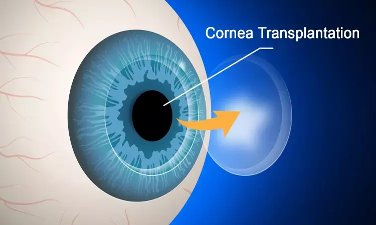
Over the years, Descemet membrane endothelial keratoplasty (DMEK) has become one of the most common techniques of corneal transplantation for the treatment of endothelial diseases such as Fuchs endothelial corneal dystrophy and pseudophakic bullous keratopathy.
Faster visual rehabilitation, improved visual outcomes, and reduced risk for rejection are among the several advantages that DMEK offers compared with penetrating keratoplasty. However, several challenges come with DMEK, such as the required skills for graft preparation, longer learning curve and management of early postoperative graft detachments.
One of the key steps in DMEK is the removal of the recipient's diseased Descemet membrane (DM) and the associated endothelial cells (descemetorhexis) before transplanting the host DM. Incomplete descemetorhexis can leave tags of DM in the recipient eye, leading to increased risk for postoperative graft detachment and preventing optimal visual outcomes in cases where these diseased DM remnants remain in the graft–host stromal interface.
Suboptimal visualization of the recipient DM during descemetorhexis can be responsible for inaccurate recipient DM removal; therefore, different methods to enhance surgical visualization of the thin recipient DM layer without increasing surgical difficulty and time have been explored. Balanced salt solution (BSS), ophthalmic viscosurgical devices (OVDs), trypan blue staining, and air in the anterior chamber (AC) have all been used during this surgical step. Air offers the best DM visualization by creating an optical interface at the endothelial layer because of the enhanced surface tension from the air–fluid interface. In addition, air offers better DM control; however, it is less popular with surgeons because of limited AC stability and the need for multiple air injections in the AC, which leads to longer surgical times.
In this study, authors Coco et al described a novel surgical technique for descemetorhexis performed under a combination of OVD and air, which offered both the advantages of improved visualization through the presence of air at the endothelial interface in addition to a stable AC and faster procedure.
TECHNIQUE
A superior stab incision with a 20-gauge micro vitreo retinal-Lance was made, and cohesive OVD with sodium hyaluronate 1% was injected into the AC through a right cannula, followed by air injection.
A reverse Sinskey hook was then inserted through the stab incision and initial scoring of the circumferential margins of the recipient DM was performed, starting from the opposite side from the stab incision.
No mark on the cornea was made to define the size of descemetorhexis; however, the aim was for approximately 8.5 to 9 mm. Gentle upward pressure was applied while scoring to tear DM without damaging the stroma.
When the scoring completed, covering approximately 360 degrees, the reverse Sinskey hook was used to bring DM edges toward the center. This allowed DM to fold onto itself and was then removed from the AC. Air at the endothelial interface provided optimal surgical visualization of the recipient DM ensured a stable AC, and provided enough pressure to allow DM to fold onto itself without flapping back, making the descemetorhexis a very controlled procedure.
The procedure finished with the removal of fine DM remnants with the 25-gauge Grieshaber MaxGrip forceps aided by the optimal DM visualization. Escape of air from the AC was not noted and, therefore, no requirement to refill the AC during the procedure. A caliper was used at the end of descemetorhexis to confirm its size. Recipient DM removal should oversize the donor tissue by at least 0.5 mm. Air and viscoelastic were then removed from the AC through an irrigation/aspiration device, and DMEK surgery was completed.
Adequate removal of recipient DM is an essential step in endothelial keratoplasty. Suboptimal DM visualization can lead to failure to remove small remnants of recipient DM during surgery. The presence of these remnants can result in inadequate adhesion of the transplanted tissue to the recipient cornea. This can lead to postoperative detachments and possible suboptimal visual results. Authors presented a new surgical technique combining the use of cohesive OVD and air in the AC for descemetorhexis, which allowed for better visualization of DM during surgery and, at the same time, offered a stable AC, making complete DM removal easier while potentially offering better surgical outcomes.
The authors concluded, "Optimal DM visualization makes this technique easy to perform even for less experienced surgeons and could potentially reduce the learning curve and improve postoperative outcomes. The only additional step was the need to remove the OVD from the AC through an irrigation/aspiration device; however, given the frequent combination of DMEK with cataract surgery, most cases should not require the use of additional instruments. We believe that this technique could offer a step forward in making DMEK surgery more accessible and accurate while reducing the risks for both detachment in the early postoperative period and suboptimal visual results because of the presence of remnants of the recipient's diseased DM."
Source: Coco et al; Cornea 2021;40:1215–1217
Dr Ishan Kataria has done his MBBS from Medical College Bijapur and MS in Ophthalmology from Dr Vasant Rao Pawar Medical College, Nasik. Post completing MD, he pursuid Anterior Segment Fellowship from Sankara Eye Hospital and worked as a competent phaco and anterior segment consultant surgeon in a trust hospital in Bathinda for 2 years.He is currently pursuing Fellowship in Vitreo-Retina at Dr Sohan Singh Eye hospital Amritsar and is actively involved in various research activities under the guidance of the faculty.
Dr Kamal Kant Kohli-MBBS, DTCD- a chest specialist with more than 30 years of practice and a flair for writing clinical articles, Dr Kamal Kant Kohli joined Medical Dialogues as a Chief Editor of Medical News. Besides writing articles, as an editor, he proofreads and verifies all the medical content published on Medical Dialogues including those coming from journals, studies,medical conferences,guidelines etc. Email: drkohli@medicaldialogues.in. Contact no. 011-43720751


