- Home
- Medical news & Guidelines
- Anesthesiology
- Cardiology and CTVS
- Critical Care
- Dentistry
- Dermatology
- Diabetes and Endocrinology
- ENT
- Gastroenterology
- Medicine
- Nephrology
- Neurology
- Obstretics-Gynaecology
- Oncology
- Ophthalmology
- Orthopaedics
- Pediatrics-Neonatology
- Psychiatry
- Pulmonology
- Radiology
- Surgery
- Urology
- Laboratory Medicine
- Diet
- Nursing
- Paramedical
- Physiotherapy
- Health news
- Fact Check
- Bone Health Fact Check
- Brain Health Fact Check
- Cancer Related Fact Check
- Child Care Fact Check
- Dental and oral health fact check
- Diabetes and metabolic health fact check
- Diet and Nutrition Fact Check
- Eye and ENT Care Fact Check
- Fitness fact check
- Gut health fact check
- Heart health fact check
- Kidney health fact check
- Medical education fact check
- Men's health fact check
- Respiratory fact check
- Skin and hair care fact check
- Vaccine and Immunization fact check
- Women's health fact check
- AYUSH
- State News
- Andaman and Nicobar Islands
- Andhra Pradesh
- Arunachal Pradesh
- Assam
- Bihar
- Chandigarh
- Chattisgarh
- Dadra and Nagar Haveli
- Daman and Diu
- Delhi
- Goa
- Gujarat
- Haryana
- Himachal Pradesh
- Jammu & Kashmir
- Jharkhand
- Karnataka
- Kerala
- Ladakh
- Lakshadweep
- Madhya Pradesh
- Maharashtra
- Manipur
- Meghalaya
- Mizoram
- Nagaland
- Odisha
- Puducherry
- Punjab
- Rajasthan
- Sikkim
- Tamil Nadu
- Telangana
- Tripura
- Uttar Pradesh
- Uttrakhand
- West Bengal
- Medical Education
- Industry
Riboflavin at 4˚C aids in Pain Management after Crosslinking for Keratoconus Patients, finds Study

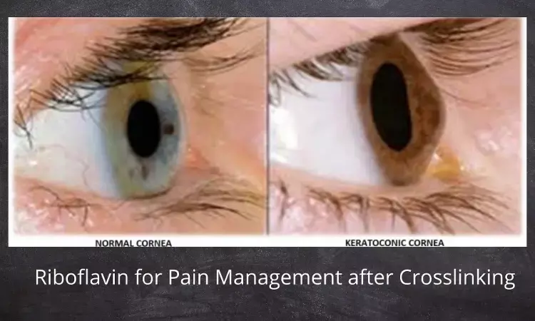
Keratoconus is a bilateral, noninflammatory, and progressive corneal ectasia that is characterized by biomechanical and biochemical instabilities of stromal collagen, leading to a decrease in corneal thickness and a variation in posterior and anterior corneal curvatures. The progressive corneal thinning leads to the characteristic cone-shaped protrusion. It is usually present during adolescence and progresses until the third or fourth decade of life.
Treatment of keratoconus can be divided into the following 2 groups:
- techniques for optical optimization and
- strengthening techniques.
Optical enhancement techniques are contact lenses, phakic lenses, or corneal transplants. Currently, the only treatment that has been shown to be effective in strengthening the cornea and halting progression is collagen cross-linking (CXL).
Pain is a major disadvantage and a common patient complaint after the CXL procedure. It may be due to damage to corneal sensory nerve fibers or locally released inflammatory mediators caused by corneal debridement. These inflammatory modulators increase the spontaneous activity of exposed nerve fibers. Pain usually starts 1 hour after treatment and increases during the next 3 to 4 hours and eventually disappears once corneal reepithelization has been completed.
Laura Toro-Giraldo et al conducted a prospective and interventional randomized clinical trial registered in the National Institutes of Health Clinical Trials. The research was conducted at the Institute of Ophthalmology "Conde de Valenciana." A total of 98 patients were randomly assigned to one of the following 2 groups: cold riboflavin (4°C) group or control group (riboflavin at room temperature).
The inclusion criteria were patients of any sex, older than 18 years of age with keratoconus diagnosis who needed management with cross-linking in both eyes because of the evidence of progression. The exclusion criteria were patients who had cross-linking without epithelial debridement, unilateral crosslinking, or any other ocular pathologies besides keratoconus and any cognitive incapacity that would make the understanding of the pain test difficult. The main outcome measures were pain, tearing, photophobia, foreign body sensation, and irritation.
Cooling has been used as management in other areas of medicine because it has been demonstrated to decrease pain and swelling, especially after burn wounds and trauma to the musculoskeletal system. This principle has been applied in corneal surgery, specifically in refractive surgery.
In general, it is believed that cooling has an effect through the decrease in prostaglandins and inflammatory mediators. It is also believed that cooling reduces corneal sensation by blocking or slowing nerve fiber conduction and that it may reduce thermal injury to the cornea. Therefore, the aim of the study was to explore corneal cooling as a method of pain management in corneal accelerated CXL.
A previously validated analog pain scale was used at 2 hours post-op (PO) and at PO days 1 through. Each patient had to indicate pain intensity in a scale of 0 to 10, knowing that 0 is no pain at all and 10 is severe and incapacitating pain. The pain scores were obtained based on pain at the moment of the interview and during the previous 24 hours.
The variables taken into account were pain, tearing, photophobia, foreign body sensation, and irritation. A blinded researcher applied all the questionnaires. The demarcation line, which can be seen since 2 weeks PO, was also recorded using the mean of 4 different points (central 1- and 2-mm rings that intersect with the horizontal line in the optical coherence tomography) to asses treatment effectivity.
At 2 hours post-op, pain in the case and control groups was 3.80 ± 3.00 and 8.08 ± 2.21 (P < 0.05). Tearing, photophobia, foreign body sensation and irritation were all reduced in case group as compared to controls (P < 0.05). A statistical significant difference was maintained in pain values on day 1 (2.79 ± 3.09 and 4.91 ± 3.27 [P < 0.05], 2 (2.54 ± 2.41 and 4.00 ± 2.43 [P , 0.05]), and 4 (0.45 ± 0.76 and 1.22 ± 1.67 [P , 0.05]).
The idea of using riboflavin and UVA as a treatment for corneal ectasias was postulated in the University of Dresden in the 1990s. It was hypothesized that the interaction between these 2 created oxygen radicals that would then activate the normal lysyl oxidase pathway ending in a photochemical crosslinking of the collagen within the corneal stroma.
The location of the cross-links at molecular level is still unknown, yet the alterations in the mechanical, physical, and chemical properties of the stroma demonstrate their existence. The process consists of an aerobic phase in which the riboflavin molecules absorb the UVA and get excited to form a triplet that interacts with the triplet oxygen species to form an active singlet oxygen that interacts with carbonyl groups of collagens to form cross-links.
In the cornea, the afferent nerve endings are divided into the following 4 groups: mechanoreceptors (20%), nociceptors (70%), cold sensing fibers (10%), and silent nociceptors.
Mechanoreceptors are fast conducting A delta fibers that respond to tactile stimuli and are responsible for the foreign body sensation; in this case, they would be triggered by blinking.
Nociceptors are slow conducting C type fibers that respond to extreme temperatures, mechanical forces, exogenous chemical stimuli, and endogenous inflammatory mediators.
The cold sensing fibers are A delta and C fibers that respond to temperatures below 33°C. The silent nociceptors are not activated by mechanical or thermal stimuli but rather when local inflammation occurs.
Therefore, corneal deepithelization leads to an inflammatory process because of direct mechanical injury of the epithelial cells and basement membrane. This process of deepithelization releases cytokines including interleukin-1, tumor necrosis factor, epithelial platelet-derived growth factor, and other collagenases and metalloproteinases.
The destruction of the basement membrane allows the contact between these cytokines with the keratocytes of the stroma, inducing apoptosis and necrosis that further perpetuates the inflammatory process through the release of cytokines. These metabolites stimulate the exposed subepithelial nerve terminations increasing pain perception.
This study demonstrated that pain and associated symptoms decreased significantly in the first interview at 2 hours PO in the cases group, and afterward, pain stayed lower than the control group at PO day 1, 2 and 4. One important limitation to this study is that there was no measurement of inflammatory mediators after the procedure to correlate with the use of cooling and pain, which could potentially support the results. These findings show an alternative to accomplishing better pain management without altering the therapeutic effects of CXL.
Source: Laura Toro-Giraldo, Norma Morales Flores, Omar Santana-Cruz et al; Cornea 2021;40:1–4
Dr Ishan Kataria has done his MBBS from Medical College Bijapur and MS in Ophthalmology from Dr Vasant Rao Pawar Medical College, Nasik. Post completing MD, he pursuid Anterior Segment Fellowship from Sankara Eye Hospital and worked as a competent phaco and anterior segment consultant surgeon in a trust hospital in Bathinda for 2 years.He is currently pursuing Fellowship in Vitreo-Retina at Dr Sohan Singh Eye hospital Amritsar and is actively involved in various research activities under the guidance of the faculty.
Dr Kamal Kant Kohli-MBBS, DTCD- a chest specialist with more than 30 years of practice and a flair for writing clinical articles, Dr Kamal Kant Kohli joined Medical Dialogues as a Chief Editor of Medical News. Besides writing articles, as an editor, he proofreads and verifies all the medical content published on Medical Dialogues including those coming from journals, studies,medical conferences,guidelines etc. Email: drkohli@medicaldialogues.in. Contact no. 011-43720751


