- Home
- Medical news & Guidelines
- Anesthesiology
- Cardiology and CTVS
- Critical Care
- Dentistry
- Dermatology
- Diabetes and Endocrinology
- ENT
- Gastroenterology
- Medicine
- Nephrology
- Neurology
- Obstretics-Gynaecology
- Oncology
- Ophthalmology
- Orthopaedics
- Pediatrics-Neonatology
- Psychiatry
- Pulmonology
- Radiology
- Surgery
- Urology
- Laboratory Medicine
- Diet
- Nursing
- Paramedical
- Physiotherapy
- Health news
- Fact Check
- Bone Health Fact Check
- Brain Health Fact Check
- Cancer Related Fact Check
- Child Care Fact Check
- Dental and oral health fact check
- Diabetes and metabolic health fact check
- Diet and Nutrition Fact Check
- Eye and ENT Care Fact Check
- Fitness fact check
- Gut health fact check
- Heart health fact check
- Kidney health fact check
- Medical education fact check
- Men's health fact check
- Respiratory fact check
- Skin and hair care fact check
- Vaccine and Immunization fact check
- Women's health fact check
- AYUSH
- State News
- Andaman and Nicobar Islands
- Andhra Pradesh
- Arunachal Pradesh
- Assam
- Bihar
- Chandigarh
- Chattisgarh
- Dadra and Nagar Haveli
- Daman and Diu
- Delhi
- Goa
- Gujarat
- Haryana
- Himachal Pradesh
- Jammu & Kashmir
- Jharkhand
- Karnataka
- Kerala
- Ladakh
- Lakshadweep
- Madhya Pradesh
- Maharashtra
- Manipur
- Meghalaya
- Mizoram
- Nagaland
- Odisha
- Puducherry
- Punjab
- Rajasthan
- Sikkim
- Tamil Nadu
- Telangana
- Tripura
- Uttar Pradesh
- Uttrakhand
- West Bengal
- Medical Education
- Industry
Pseudoaneurysm of the Popliteal Artery After Revision TKA: a case report

Netherlands: Pseudo aneurysms of the popliteal artery are a rare complication after total knee arthroplasty (TKA), with reported incidence varying between 0.0095% and 0.088%. A pseudo aneurysm, or false aneurysm, is a contained local hematoma in direct connection with an artery due to a defect in the layers of the arterial vessel wall.
This is in contrast to a true aneurysm where all layers of the vessel wall are intact. Often, a pseudo aneurysm of the popliteal artery presents similar to a deep venous thrombosis (DVT): progressive swelling, pain, and tightness of the calf. If not recognized early, it has the potential to developing compartment syndrome or irreversible neurological deficits.
B.A. Schermer et al. presents a case of 66-year-old female patient with a unicompartmental knee arthroplasty who underwent conversion to a TKA because of anteroposterior instability symptoms, which she developed after a fall from the stairs 1.5 years before the revision. The patient's medical history includes asthma, hypertension, type 2 diabetes mellitus, and obesity (body mass index of 36 kg/m2).
Preoperative physical examination showed a grade 3 positive Lachman test, suggesting an anterior cruciate ligament rupture and a positive posterior sag sign indicative of concomitant posterior cruciate ligament damage. Varus and valgus stress tests showed no signs of instability; neurological examination was without abnormalities.
During surgery, a cemented revision TKA with a short-stemmed tibial component was placed. The surgery was performed without intraoperative complications, and no tourniquet was used. Furthermore, local infiltration anesthesia was applied in the posterior and anterior capsules and subcutaneously.
On postoperative day 2, the patient experienced pain in the calf and the knee and decreased sensibility of the lateral edge of the foot. Physical examination showed a tense calf and foot (not red, not shining). Palpation of the whole leg was painful, including the calf. Moreover, there was decreased sensibility of the dorsal side of toes 3-5 and over the lateral edge of the foot, which the patient had noticed after spinal anesthesia had worn off. All motor functions were intact.
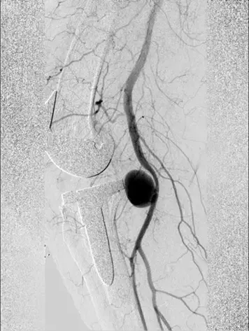
On postoperative day 4, a hematoma of the operative leg was noted. The clinical presentation seemed to best fit an expanding hematoma. Because of hindering symptoms and to exclude a DVT, a venous Duplex ultrasound of the lower leg was performed on the fourth postoperative day. On the ultrasound, no DVT was identified. However, a globular abnormality in direct relation to the popliteal artery with arterial pulsations was found. Computed tomography angiography (CTA) confirmed the presence of a pseudoaneurysm at the level of the joint space of the knee prosthesis originating from the popliteal artery with a diameter of approximately 22 mm. The popliteal artery itself was patent. Owing to a wide neck of the pseudoaneurysm (1-2 cm), neither thrombin injection nor coiling was possible given the risk of distal embolization. Also, there is a chance that bleeding might persist after coiling of the pseudoaneurysm.
Thereupon, after consultation with the interventional radiologist and the vascular surgeon, it was decided to treat the pseudoaneurysm endovascularly with a covered stent. Through an antegrade sheath in the common femoral artery, a 750-mm covered stent was placed; subsequent angiography showed complete elimination of the pseudoaneurysm. The procedure was uncomplicated, and postprocedural dual antiplatelet therapy was started for 6 months.
On day 7, the patient was discharged in good condition, with residual symptoms of pain, swelling, and hypesthesia of the dorsal side of digits 3 to 5. The stent was patent at control Duplex ultrasound after 2 weeks.
At 8 months after surgery, the patient showed good, yet slower than normal, functional recovery; the complaints of hypesthesia of digits 3 to 5 of the right foot and heel persisted.
Learning points:
- Pseudoaneurysm of the popliteal artery is a rare complication of knee arthroplasty.s
- Indirect trauma with local manipulation intraoperatively is considered as a trauma mechanism.
- The clinical presentation is similar to that of a DVT, a pulsatile mass may be distinctive.
- Often observed coincidentally on ultrasound, CTA may be per formed additionally for confirmation of diagnosis and planning of treatment.
- Usually, there is a delay in diagnosis: median intervals of 2 to 6 weeks after surgery.
- In the past, most cases were repaired with open surgery. Nowadays, endovascular intervention is preferred.
- Residual symptoms are not uncommon: hypesthesia, (neuropathic) pain, and/or swelling
Key Words: Pseudoaneurysm, Popliteal artery, Total knee arthroplasty, Revision knee arthroplasty, stent
Further reading:
Pseudoaneurysm of the Popliteal Artery After Revision Knee Arthroplasty
Biko A. Schermer, Arne C. Berger, Wouter Stomp, Joris C.T. van der Lugt.
Arthroplasty Today 13 (2022) 1- 6
https://doi.org/10.1016/j.artd.2021.11.002
MBBS, Dip. Ortho, DNB ortho, MNAMS
Dr Supreeth D R (MBBS, Dip. Ortho, DNB ortho, MNAMS) is a practicing orthopedician with interest in medical research and publishing articles. He completed MBBS from mysore medical college, dip ortho from Trivandrum medical college and sec. DNB from Manipal Hospital, Bengaluru. He has expirence of 7years in the field of orthopedics. He has presented scientific papers & posters in various state, national and international conferences. His interest in writing articles lead the way to join medical dialogues. He can be contacted at editorial@medicaldialogues.in.
Dr Kamal Kant Kohli-MBBS, DTCD- a chest specialist with more than 30 years of practice and a flair for writing clinical articles, Dr Kamal Kant Kohli joined Medical Dialogues as a Chief Editor of Medical News. Besides writing articles, as an editor, he proofreads and verifies all the medical content published on Medical Dialogues including those coming from journals, studies,medical conferences,guidelines etc. Email: drkohli@medicaldialogues.in. Contact no. 011-43720751


