- Home
- Medical news & Guidelines
- Anesthesiology
- Cardiology and CTVS
- Critical Care
- Dentistry
- Dermatology
- Diabetes and Endocrinology
- ENT
- Gastroenterology
- Medicine
- Nephrology
- Neurology
- Obstretics-Gynaecology
- Oncology
- Ophthalmology
- Orthopaedics
- Pediatrics-Neonatology
- Psychiatry
- Pulmonology
- Radiology
- Surgery
- Urology
- Laboratory Medicine
- Diet
- Nursing
- Paramedical
- Physiotherapy
- Health news
- Fact Check
- Bone Health Fact Check
- Brain Health Fact Check
- Cancer Related Fact Check
- Child Care Fact Check
- Dental and oral health fact check
- Diabetes and metabolic health fact check
- Diet and Nutrition Fact Check
- Eye and ENT Care Fact Check
- Fitness fact check
- Gut health fact check
- Heart health fact check
- Kidney health fact check
- Medical education fact check
- Men's health fact check
- Respiratory fact check
- Skin and hair care fact check
- Vaccine and Immunization fact check
- Women's health fact check
- AYUSH
- State News
- Andaman and Nicobar Islands
- Andhra Pradesh
- Arunachal Pradesh
- Assam
- Bihar
- Chandigarh
- Chattisgarh
- Dadra and Nagar Haveli
- Daman and Diu
- Delhi
- Goa
- Gujarat
- Haryana
- Himachal Pradesh
- Jammu & Kashmir
- Jharkhand
- Karnataka
- Kerala
- Ladakh
- Lakshadweep
- Madhya Pradesh
- Maharashtra
- Manipur
- Meghalaya
- Mizoram
- Nagaland
- Odisha
- Puducherry
- Punjab
- Rajasthan
- Sikkim
- Tamil Nadu
- Telangana
- Tripura
- Uttar Pradesh
- Uttrakhand
- West Bengal
- Medical Education
- Industry
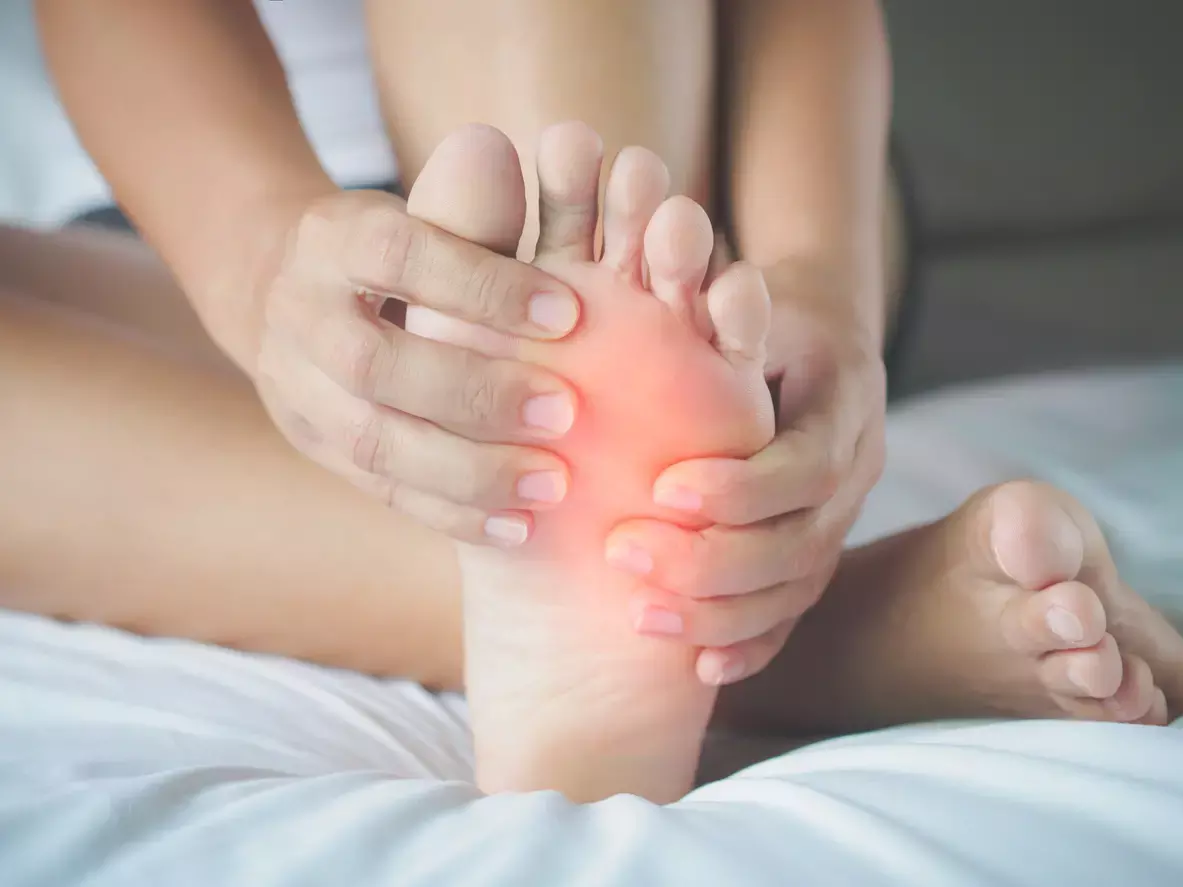
Rare case of Calcifying fibrous tumor of foot: A report

Calcifying fibrous tumor (CFT) is a rare fibroblastic tumor with distinctive histological presentation that shows benign characteristics. CFT is most commonly found in the gastrointestinal tract, the pleura, abdominal cavity, and the neck of young patients. Rare cases have been reported at other somatic soft tissue locations as well as in older adults.
Upasana Upadhyay Bharadwaj et al reported a case of a 25-year-old man with documented imaging findings of calcifying fibrous tumor in the distal lower extremity in ‘Skeletal Radiology’ journal.
Case report
A 25-year-old man presented with a right foot mass that was first noted 4 years earlier. A medical history of depression, loss of appetite, weight loss, acute kidney injury, sciatica, and closed reduction percutaneous pinning of the left hand was reported. Medical attention was sought due to increasing pain in the right foot and concern that the mass may have grown in size. The patient had no limitations in activity but reported worsening discomfort while walking.
An anteroposterior radiograph of the right foot obtained at initial consultation showed a large soft tissue opacity in the medial aspect of the foot with scattered punctate, amorphous calcifications. Based on the clinical presentation and initial imaging, differential diagnosis included low-grade chondrosarcoma, synovial sarcoma, and myositis ossificans, while no definite bony abnormality was noted the lesion was calcified and showed ring-and-arc-like mineralization, which were found to be suggestive of extra-skeletal low-grade chondrosarcoma given the long disease history.
Contrast-enhanced 3.0 T magnetic resonance imaging (MRI) revealed a 6.5 cm×2.5 cm×8.5 cm mass hypointense on T1- and T2-weighted images, which demonstrated heterogenous low-grade enhancement on fat-suppressed contrast enhanced T1-weighted images on the medial plantar aspect of the right midfoot.MR imaging features were suggestive of a fibrotic lesion.
An initial core biopsy demonstrated a hypocellular fibrous lesion with scattered calcifications. The subsequent excision specimen consisted of multiple irregular fragments of white-tan firm tissue with a gritty cut surface. Microscopically, the lesion consisted of a multinodular mass composed of dense collagen and widely dispersed calcifications, which varied from irregularly shaped to psammomatous. The lesion was sparsely cellular with cytologically bland bipolar spindle cells dispersed in the collagen and occasional lymphocytes and plasma cells in a perivascular distribution. By immunohistochemistry, the tumor was patchy positive for CD34 but negative for STAT6, epithelial membrane antigen, S100 protein, Pankeratin, Desmin, smooth muscle actin, MUC4, nuclear beta-catenin, and ALK1. Findings were overall consistent with calcifying fibrous tumor (CFT).
Clinical follow up Based on the initial core biopsy findings favoring CFT, the patient was offered surgical resection but opted to observe. As the foot continued to be painful and swelling increased in size, the patient requested surgical resection 1 year later.
The authors commented – “In conclusion, CFT is a rare tumor outside the abdominal and thoracic cavities but should be considered as a differential diagnosis in the case of calcifying soft tissue mass at other anatomic locations. Low signal intensity with low grade enhancement on MRI as well as indolent course and stable prognosis could prompt its diagnosis even in less common locations.”
Further reading:
Calcifying fibrous tumor: a rare case in the foot
Upasana Upadhyay Bharadwaj, Zehra Akkaya et al
Skeletal Radiology (2023) 52:1619–1623
https://doi.org/10.1007/s00256-023-04282-y
MBBS, Dip. Ortho, DNB ortho, MNAMS
Dr Supreeth D R (MBBS, Dip. Ortho, DNB ortho, MNAMS) is a practicing orthopedician with interest in medical research and publishing articles. He completed MBBS from mysore medical college, dip ortho from Trivandrum medical college and sec. DNB from Manipal Hospital, Bengaluru. He has expirence of 7years in the field of orthopedics. He has presented scientific papers & posters in various state, national and international conferences. His interest in writing articles lead the way to join medical dialogues. He can be contacted at editorial@medicaldialogues.in.
Dr Kamal Kant Kohli-MBBS, DTCD- a chest specialist with more than 30 years of practice and a flair for writing clinical articles, Dr Kamal Kant Kohli joined Medical Dialogues as a Chief Editor of Medical News. Besides writing articles, as an editor, he proofreads and verifies all the medical content published on Medical Dialogues including those coming from journals, studies,medical conferences,guidelines etc. Email: drkohli@medicaldialogues.in. Contact no. 011-43720751


