- Home
- Medical news & Guidelines
- Anesthesiology
- Cardiology and CTVS
- Critical Care
- Dentistry
- Dermatology
- Diabetes and Endocrinology
- ENT
- Gastroenterology
- Medicine
- Nephrology
- Neurology
- Obstretics-Gynaecology
- Oncology
- Ophthalmology
- Orthopaedics
- Pediatrics-Neonatology
- Psychiatry
- Pulmonology
- Radiology
- Surgery
- Urology
- Laboratory Medicine
- Diet
- Nursing
- Paramedical
- Physiotherapy
- Health news
- Fact Check
- Bone Health Fact Check
- Brain Health Fact Check
- Cancer Related Fact Check
- Child Care Fact Check
- Dental and oral health fact check
- Diabetes and metabolic health fact check
- Diet and Nutrition Fact Check
- Eye and ENT Care Fact Check
- Fitness fact check
- Gut health fact check
- Heart health fact check
- Kidney health fact check
- Medical education fact check
- Men's health fact check
- Respiratory fact check
- Skin and hair care fact check
- Vaccine and Immunization fact check
- Women's health fact check
- AYUSH
- State News
- Andaman and Nicobar Islands
- Andhra Pradesh
- Arunachal Pradesh
- Assam
- Bihar
- Chandigarh
- Chattisgarh
- Dadra and Nagar Haveli
- Daman and Diu
- Delhi
- Goa
- Gujarat
- Haryana
- Himachal Pradesh
- Jammu & Kashmir
- Jharkhand
- Karnataka
- Kerala
- Ladakh
- Lakshadweep
- Madhya Pradesh
- Maharashtra
- Manipur
- Meghalaya
- Mizoram
- Nagaland
- Odisha
- Puducherry
- Punjab
- Rajasthan
- Sikkim
- Tamil Nadu
- Telangana
- Tripura
- Uttar Pradesh
- Uttrakhand
- West Bengal
- Medical Education
- Industry
Rare case of intracortical chondroblastoma of the diaphysis in skeletally mature patient reported

Chondroblastomas characteristically occur in skeletally immature patients, and arise within the medullary canal of the epiphysis. Madeline A. Sauer et al report a rare case of an intracortical chondroblastoma arising in the diaphysis, and occurring in an adult in his 3rd decade of life. Immunohistochemistry results were critical to confirmation of this rare diagnosis, with immunohistochemistry showing S100, DOG1, and H3K36me3 positivity in the neoplastic cells.
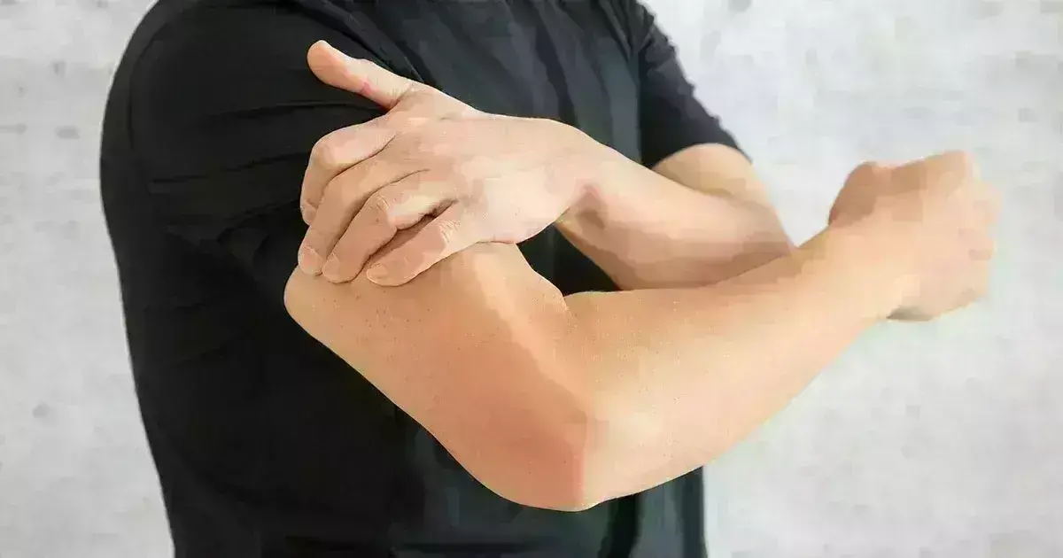
A 27-year-old right-hand dominant male presented to the orthopedic clinic with a 2-month history of persistent 2/10 right shoulder pain. He denied any history of trauma or injury to the arm. The pain was exacerbated by activity especially abduction. Lifting any weight over 10 pounds caused shoulder pain, but the patient had no pain with forward flexion. Physical exam at the time noted tenderness along the distal and lateral aspect of the deltoid near its insertion into the humerus without gross atrophy, masses, or deformity. Impingement and rotator cuff tests were negative. Radiographs and MRI were obtained, and the patient was referred to orthopedic oncology. A biopsy was then obtained.
Radiographs showed a sharply demarcated, expansile lesion arising in the cortex of the mid humerus. A thin shell of periosteal new bone surrounded the external surface. Faint calcifications within the lesion could represent either osteoid or cartilage calcifications. Differential diagnosis at this point was periosteal chondroma, intracortical osteoblastoma, or intracortical chondroma.
MRI showed the lesion was low signal intensity on T1 weighted images, with struts of signal void suggesting residual bone. On fluid sensitive images, much of the lesion remained low signal intensity, with small, rounded, well-defined areas of high signal intensity. There were patchy areas of contrast enhancement.
Initial biopsy showed scattered groups of round to ovoid cells with occasional multinucleated giant cells. The findings were indicative of a giant cell lesion with a differential of giant cell tumor vs. chondroblastoma. Immunohistochemistry demonstrated the neoplastic cells to be strongly positive for S100 protein, DOG1, and H3K36me3. The strong reaction with DOG1 and H3K36me3 supported the diagnosis of chondroblastoma.
The patient initially declined surgery but underwent curettage & microparticulate bone grafting 5 months later. The surgical specimen at that time showed sheets of small, uniformly round to oval cells associated with aggregates of chondroid. The individual cells had scant to modest amounts of cytoplasm surrounding the nuclei. The individual nuclei were round to oval in shape often with nuclear membrane folds or creases (arrow), consistent with chondroblastoma. Scattered among the mononuclear cells were multinucleated giant osteoclast-like giant cells having between 3 and 10 nuclei. Scattered mitoses were present but uncommon.
Because of the risk of local recurrence of chondroblastoma, adjuvant treatment was also administered with intra-medullary phenol. The patient was asymptomatic at the 6-week follow-up clinical visit.
The authors commented "because of the rarity of diaphyseal chondroblastomas, the occurrence of a neoplasm with the classic histomorphologic appearance of a chondroblastoma in this location should prompt ancillary testing including immunohistochemistry and mutational analysis."
Further reading:
A report of an intracortical chondroblastoma of the diaphysis in a skeletally mature patient
Madeline A. Sauer, Paul Stegelmeier et al
Skeletal Radiology
https://doi.org/10.1007/s00256-022-04088-4
MBBS, Dip. Ortho, DNB ortho, MNAMS
Dr Supreeth D R (MBBS, Dip. Ortho, DNB ortho, MNAMS) is a practicing orthopedician with interest in medical research and publishing articles. He completed MBBS from mysore medical college, dip ortho from Trivandrum medical college and sec. DNB from Manipal Hospital, Bengaluru. He has expirence of 7years in the field of orthopedics. He has presented scientific papers & posters in various state, national and international conferences. His interest in writing articles lead the way to join medical dialogues. He can be contacted at editorial@medicaldialogues.in.
Dr Kamal Kant Kohli-MBBS, DTCD- a chest specialist with more than 30 years of practice and a flair for writing clinical articles, Dr Kamal Kant Kohli joined Medical Dialogues as a Chief Editor of Medical News. Besides writing articles, as an editor, he proofreads and verifies all the medical content published on Medical Dialogues including those coming from journals, studies,medical conferences,guidelines etc. Email: drkohli@medicaldialogues.in. Contact no. 011-43720751


