- Home
- Medical news & Guidelines
- Anesthesiology
- Cardiology and CTVS
- Critical Care
- Dentistry
- Dermatology
- Diabetes and Endocrinology
- ENT
- Gastroenterology
- Medicine
- Nephrology
- Neurology
- Obstretics-Gynaecology
- Oncology
- Ophthalmology
- Orthopaedics
- Pediatrics-Neonatology
- Psychiatry
- Pulmonology
- Radiology
- Surgery
- Urology
- Laboratory Medicine
- Diet
- Nursing
- Paramedical
- Physiotherapy
- Health news
- Fact Check
- Bone Health Fact Check
- Brain Health Fact Check
- Cancer Related Fact Check
- Child Care Fact Check
- Dental and oral health fact check
- Diabetes and metabolic health fact check
- Diet and Nutrition Fact Check
- Eye and ENT Care Fact Check
- Fitness fact check
- Gut health fact check
- Heart health fact check
- Kidney health fact check
- Medical education fact check
- Men's health fact check
- Respiratory fact check
- Skin and hair care fact check
- Vaccine and Immunization fact check
- Women's health fact check
- AYUSH
- State News
- Andaman and Nicobar Islands
- Andhra Pradesh
- Arunachal Pradesh
- Assam
- Bihar
- Chandigarh
- Chattisgarh
- Dadra and Nagar Haveli
- Daman and Diu
- Delhi
- Goa
- Gujarat
- Haryana
- Himachal Pradesh
- Jammu & Kashmir
- Jharkhand
- Karnataka
- Kerala
- Ladakh
- Lakshadweep
- Madhya Pradesh
- Maharashtra
- Manipur
- Meghalaya
- Mizoram
- Nagaland
- Odisha
- Puducherry
- Punjab
- Rajasthan
- Sikkim
- Tamil Nadu
- Telangana
- Tripura
- Uttar Pradesh
- Uttrakhand
- West Bengal
- Medical Education
- Industry
EULAR releases guidelines on imaging use in the clinical management of crystal-induced arthropathies

Austria: The European Alliance of Associations for Rheumatology (EULAR) has released recommendations on imaging for diagnosing and managing crystal-induced arthropathies (CiAs) in clinical practice.
The purpose of these guidelines, published in Annals of the Rheumatic Diseases, is to help clinicians when applying imaging techniques for all common CiAs and include the full spectrum of imaging in clinical practice, including monitoring disease activity, diagnosis, treatment response, and the prediction of outcome. These guidelines are also useful for healthcare providers when making decisions regarding management in terms of imagining this patient population.
"These are the first recommendations that encompass the major forms of CiA and guide the use of common imaging modalities in the clinical management of this disease group," wrote an international team comprising 25 rheumatologists, radiologists, methodologists, healthcare professionals, and patient research partners from 11 countries.
During the development of these guidelines, the team considered the heterogeneous clinical phenotype of these diseases. Five overarching principles and 10 recommendations were developed encompassing the role of imaging in various aspects of patient management: making a CiA diagnosis, monitoring inflammation and damage, predicting outcomes, response to treatment, guided interventions and patient education. Overall, the LoA for the recommendations was high.
Five statements are as follows:
- Imaging in CiAs provides useful information on crystal deposition, inflammation, and structural damage.
- The presence of imaging abnormalities, particularly those related to crystal deposition, may not always be related to clinical manifestations.
- CiAs are typically characterized by intermittent, acute episodes of inflammation. However, they may also exhibit a persistent disease course with or without superimposed flares.
- Imaging in CiAs should be performed and interpreted by trained healthcare professionals.
- Patient information, including medical history, physical and laboratory examination, synovial fluid and tissue analysis, should be taken into account when considering imaging in CiAs.
The following recommendations were developed:
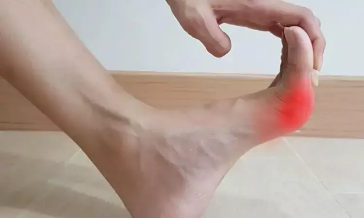
- When conducting imaging in CiAs, both symptomatic areas and disease-specific target sites, such as first metatarsophalangeal in gout, knee and wrist in calcium pyrophosphate deposition (CPPD), shoulder in basic calcium phosphate deposition (BCPD), should be considered.
- During the diagnostic assessment of gout, ultrasound and dual-energy computed tomography (DECT) are recommended imaging modalities.
- When characteristic features of monosodium urate (MSU) crystal deposition on ultrasound, such as double-contour sign or tophi, or on DECT are identified, synovial fluid analysis is not needed to confirm a diagnosis of gout.
- During the diagnostic assessment of CPPD, conventional radiography (CR) and ultrasound (or CT if axial involvement is suspected) are recommended imaging modalities.
- During the diagnostic assessment of BCPD, imaging is necessary. CR or ultrasound is the recommended modality.
- In CPPD and BCPD, serial imaging is not recommended unless there is an unexpected change in clinical characteristics.
- In gout, ultrasound and DECT can be used to monitor crystal deposition and in the case of ultrasound, also inflammation. Both modalities provide additional information in addition to clinical and biochemical assessment. CR can be used to assess structural damage due to gout when ultrasound/DECT is unavailable. The decision on when to repeat imaging depends on the clinical circumstances.
- If synovial fluid analysis is required in CiAs assessment, ultrasound guidance should be used in cases where aspiration based on anatomical landmarks is challenging.
- In gout, assessing the amount of MSU crystal deposition by ultrasound or DECT may be used to predict future flares.
- Showing and explaining imaging findings of CiAs to people with such conditions may help them understand their condition and improve treatment adherence in gout.
The authors encourage future research to investigate which imaging techniques provide the best early and accurate CiAs diagnosis and determine the utility of ultrasound and DECT in monitoring crystal deposition and inflammation.
They are also interested in the diagnostic performance of emerging advanced imaging techniques in this patient population and want to assess the utility of presenting and discussing imaging findings with patients with crystal-induced arthropathy concerning disease management.
Reference:
Mandl P, D’Agostino MA, Navarro-Compán V, et al2023 EULAR recommendations on imaging in diagnosis and management of crystal-induced arthropathies in clinical practiceAnnals of the Rheumatic Diseases Published Online First: 06 February 2024. doi: 10.1136/ard-2023-224771
Dr Kamal Kant Kohli-MBBS, DTCD- a chest specialist with more than 30 years of practice and a flair for writing clinical articles, Dr Kamal Kant Kohli joined Medical Dialogues as a Chief Editor of Medical News. Besides writing articles, as an editor, he proofreads and verifies all the medical content published on Medical Dialogues including those coming from journals, studies,medical conferences,guidelines etc. Email: drkohli@medicaldialogues.in. Contact no. 011-43720751


