
Advanced Imaging Enables Earlier Detection of Joint Inflammation in kids, finds research
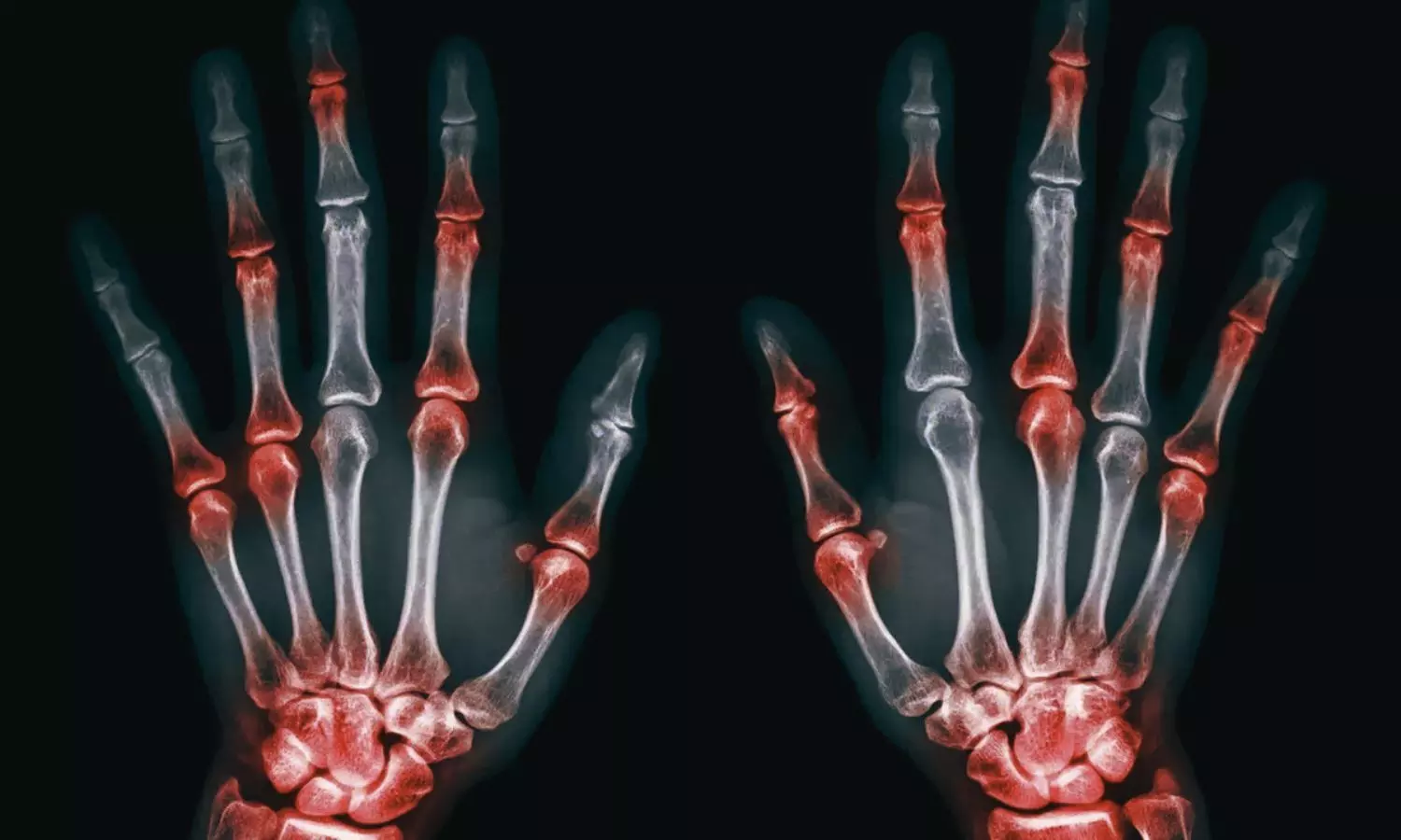


A recent comprehensive review published in the Indian Journal of Radiology and Imaging in August 2025 highlights a major clinical shift from traditional X-rays to advanced, highly sensitive imaging techniques. Driven by modern anti-inflammatory treatments, this crucial transition empowers clinicians to catch inflammation early and stop irreversible joint damage before it starts.
While physical exams for Juvenile Idiopathic Arthritis (JIA) often miss early or hidden joint damage, advanced imaging effectively bridges this diagnostic gap. To address this, researchers from AIIMS comprehensively reviewed how these modern imaging tools accurately monitor disease activity and treatment responses in children.
Therefore, the comprehensive review compared the effectiveness of traditional X-rays against ultrasound and magnetic resonance imaging (MRI) for diagnosing and monitoring pediatric joint diseases. Additionally, it highlighted key imaging-based scoring systems to help clinicians accurately evaluate disease severity in unclear cases.
Key Clinical Findings of the Review Includes:
• Radiographic Limitations: Reviewers noted that while conventional radiographs remain widely utilized as a preliminary investigation, they lack the sensitivity to detect early inflammatory changes, instead predominantly revealing advanced disease sequelae such as late-stage joint space reduction and bony ankylosis.
• Sonographic Efficacy: Investigators highlighted that ultrasonography offers a superior, radiation-free alternative to physical examinations for detecting synovial hypervascularity and subclinical synovitis, proving especially valuable for guiding targeted intra-articular therapeutic injections with minimal complications.
• MRI Superiority: Researchers affirmed that MRI currently stands as the most sensitive modality for comprehensively identifying early bone marrow edema, active synovial inflammation, and osteochondral damage well before permanent morphological destruction appears on standard X-rays.
• Comprehensive Screening: Scientists emphasized that whole-body MRI allows for the effective simultaneous quantification of disease burden across multiple joint sites in a single session, emerging as the strongest predictor of disease relapse in clinically asymptomatic patients.
• Distinguishing Mimics: Authors indicated that advanced cross-sectional imaging features, specifically distinct bone marrow edema locations and synovial tissue patterns, are indispensable for reliably differentiating monoarticular JIA from close infectious mimics like tubercular arthritis.
The results suggest that integrating sensitive, non-ionizing imaging technologies into routine diagnostic pathways fundamentally enhances the ability of clinicians to detect subclinical inflammation early in the disease course. Furthermore, leveraging comprehensive MRI-based scoring systems empowers physicians to meticulously monitor therapeutic efficacy and halt insidious disease progression before permanent functional impairment occurs.
Thus, the review concludes that healthcare professionals should consider incorporating advanced, soft-tissue-sensitive imaging evaluations into their standard pediatric workflows to better detect clinically silent inflammation and carefully tailor individual early intervention strategies.
Although inherent limitations exist, such as unavoidable operator dependence in sonographic evaluations and an absence of formally endorsed scoring criteria for spinal involvement, there remains a mild necessity for future research directed toward establishing globally standardized whole-body imaging protocols that can minimize acquisition times while maximizing diagnostic accuracy.
Reference
Chandola, S., Bagri, N., & Jana, M. (2025). Imaging in Juvenile Idiopathic Arthritis: A Comprehensive Review. Indian Journal of Radiology and Imaging, 36(3), 426–439.