- Home
- Medical news & Guidelines
- Anesthesiology
- Cardiology and CTVS
- Critical Care
- Dentistry
- Dermatology
- Diabetes and Endocrinology
- ENT
- Gastroenterology
- Medicine
- Nephrology
- Neurology
- Obstretics-Gynaecology
- Oncology
- Ophthalmology
- Orthopaedics
- Pediatrics-Neonatology
- Psychiatry
- Pulmonology
- Radiology
- Surgery
- Urology
- Laboratory Medicine
- Diet
- Nursing
- Paramedical
- Physiotherapy
- Health news
- Fact Check
- Bone Health Fact Check
- Brain Health Fact Check
- Cancer Related Fact Check
- Child Care Fact Check
- Dental and oral health fact check
- Diabetes and metabolic health fact check
- Diet and Nutrition Fact Check
- Eye and ENT Care Fact Check
- Fitness fact check
- Gut health fact check
- Heart health fact check
- Kidney health fact check
- Medical education fact check
- Men's health fact check
- Respiratory fact check
- Skin and hair care fact check
- Vaccine and Immunization fact check
- Women's health fact check
- AYUSH
- State News
- Andaman and Nicobar Islands
- Andhra Pradesh
- Arunachal Pradesh
- Assam
- Bihar
- Chandigarh
- Chattisgarh
- Dadra and Nagar Haveli
- Daman and Diu
- Delhi
- Goa
- Gujarat
- Haryana
- Himachal Pradesh
- Jammu & Kashmir
- Jharkhand
- Karnataka
- Kerala
- Ladakh
- Lakshadweep
- Madhya Pradesh
- Maharashtra
- Manipur
- Meghalaya
- Mizoram
- Nagaland
- Odisha
- Puducherry
- Punjab
- Rajasthan
- Sikkim
- Tamil Nadu
- Telangana
- Tripura
- Uttar Pradesh
- Uttrakhand
- West Bengal
- Medical Education
- Industry
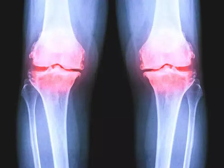
Are Weight-Bearing Knee Digital Radiographs better for assessment of Severity of OA?

Tamilnadu, India: Weight-bearing radiographs are preferred for joint space width (JSW) assessment in OA , sometimes non-weightbearing (NWB) radiographs are also done.
Patients above 50 years old with symptoms (knee pain, functional limitations, and crepitus) were included in the study. Expert excluded patients with post-surgical evaluation, trauma, and infection. JSW and Q angle measurements with respect to weight-bearing and non-weight-bearing positions were made by the same medical expert to avoid inter-observer variation.
The observations of the study were:
• Medial knee JSW varies from 0.49 to 5.75 mm in weight-bearing radiograph and 0.53 mm to 6.16 mm in nonweight-bearing radiograph.
• Q angle varies from 8.72° to 14.58° in weight-bearing position and 10.13° to 14.92° in nonweight-bearing position.
• Q angle (<11.72°) and medial JSW (<3.275) were found to be significantly associated with the rapid progression of knee varus OA (p<0.001).
The authors concluded that - Outcomes with respect to weight-bearing and non-weight bearing radiographs/positions vary significantly. For evaluating varus deformity, weight-bearing radiographs are necessary. Non-weight-bearing radiographs appear to exaggerate the deformation in early varus deformity, although they appear to underestimate it in advanced deformation. The result of the study shows that knee mJSW measurements from digital radiographs are relatively the same for mild and severe cases than moderate to severe cases in both WB and NWB positions. From the study, we found that there is no significant JSW difference between non-weight-bearing and weight-bearing radiographs in the initial stages of OA but patients are unable to put load due to pain. Therefore, using non-weight-bearing radiographs rather than weight bearing radiographs to analyze knee joint space width is very helpful in the initial stages of OA. We noticed a tendency to increase joint space width and external rotation of the tibia during weight-bearing position. This result is particularly significant considering that the radiographic positions are the important factors for the diagnosis of OA. These findings show that weight-bearing knee radiographs are useful to track the progression (severity) of knee varus OA than non-weight-bearing radiographs. Patients whose Q angle and knee JSW measures are below the limitations need more attention.
Further reading:
Do Weight Bearing Knee Digital Radiographs Help to Track the Severity of OA?
S. Sheik Abdullah, M. Pallikonda Rajasekaran.
Indian Journal of Orthopaedics (2022) 56:664–671
https://doi.org/10.1007/s43465-021-00560-w
MBBS, Dip. Ortho, DNB ortho, MNAMS
Dr Supreeth D R (MBBS, Dip. Ortho, DNB ortho, MNAMS) is a practicing orthopedician with interest in medical research and publishing articles. He completed MBBS from mysore medical college, dip ortho from Trivandrum medical college and sec. DNB from Manipal Hospital, Bengaluru. He has expirence of 7years in the field of orthopedics. He has presented scientific papers & posters in various state, national and international conferences. His interest in writing articles lead the way to join medical dialogues. He can be contacted at editorial@medicaldialogues.in.
Dr Kamal Kant Kohli-MBBS, DTCD- a chest specialist with more than 30 years of practice and a flair for writing clinical articles, Dr Kamal Kant Kohli joined Medical Dialogues as a Chief Editor of Medical News. Besides writing articles, as an editor, he proofreads and verifies all the medical content published on Medical Dialogues including those coming from journals, studies,medical conferences,guidelines etc. Email: drkohli@medicaldialogues.in. Contact no. 011-43720751


