- Home
- Medical news & Guidelines
- Anesthesiology
- Cardiology and CTVS
- Critical Care
- Dentistry
- Dermatology
- Diabetes and Endocrinology
- ENT
- Gastroenterology
- Medicine
- Nephrology
- Neurology
- Obstretics-Gynaecology
- Oncology
- Ophthalmology
- Orthopaedics
- Pediatrics-Neonatology
- Psychiatry
- Pulmonology
- Radiology
- Surgery
- Urology
- Laboratory Medicine
- Diet
- Nursing
- Paramedical
- Physiotherapy
- Health news
- Fact Check
- Bone Health Fact Check
- Brain Health Fact Check
- Cancer Related Fact Check
- Child Care Fact Check
- Dental and oral health fact check
- Diabetes and metabolic health fact check
- Diet and Nutrition Fact Check
- Eye and ENT Care Fact Check
- Fitness fact check
- Gut health fact check
- Heart health fact check
- Kidney health fact check
- Medical education fact check
- Men's health fact check
- Respiratory fact check
- Skin and hair care fact check
- Vaccine and Immunization fact check
- Women's health fact check
- AYUSH
- State News
- Andaman and Nicobar Islands
- Andhra Pradesh
- Arunachal Pradesh
- Assam
- Bihar
- Chandigarh
- Chattisgarh
- Dadra and Nagar Haveli
- Daman and Diu
- Delhi
- Goa
- Gujarat
- Haryana
- Himachal Pradesh
- Jammu & Kashmir
- Jharkhand
- Karnataka
- Kerala
- Ladakh
- Lakshadweep
- Madhya Pradesh
- Maharashtra
- Manipur
- Meghalaya
- Mizoram
- Nagaland
- Odisha
- Puducherry
- Punjab
- Rajasthan
- Sikkim
- Tamil Nadu
- Telangana
- Tripura
- Uttar Pradesh
- Uttrakhand
- West Bengal
- Medical Education
- Industry
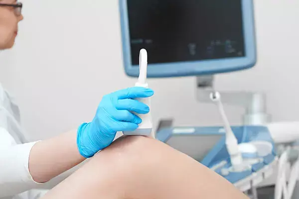
Dynamic ultrasound may quantitatively assess medial patellofemoral complex injury in multiple clinical settings

The use of imaging to diagnose patellofemoral instability is often limited by the inability to dynamically load the joint during assessment. Therefore, the diagnosis is typically based on physical examination using the glide test to assess and quantify lateral patellar translation. However, precise quantification with this technique remains difficult.
Bhimani Et al found in a Controlled laboratory study that it is possible to quantify patellar position using ultrasound imaging under dynamic loading conditions to distinguish between knees with and without medial patellofemoral complex (MPFC) injury. Investigation was performed at Massachusetts General Hospital, Harvard Medical School, Boston, Massachusetts, USA
In 10 cadaveric knees, the medial patellofemoral distance was measured to quantify patellar position from 00 to 400 of knee flexion at 100 increments. Knees were evaluated at each flexion angle under unloaded conditions and with 20 N of laterally directed force on the patella to mimic the glide test. Patellar position measurements were made on ultrasound images obtained before and after MPFC transection and compared for significant differences.
To determine the ability of medial patellofemoral measurements to differentiate between MPFC-intact and MPFC-deficient states, area under the receiver operating characteristic (ROC) curve analysis and the Delong test were used.
The optimal cutoff value to distinguish between the deficient and intact states was determined using the Youden J statistic.
The results of the study were -
• A significant increase in medial patellofemoral distance was observed in the MPFC-deficient state as compared with the intact state at all flexion angles (P =.005 to P < .001).
• When compared with the intact state, MPFC deficiency increased medial patellofemoral distance by 32.8% (6 mm) at 200 of knee flexion under 20-N load.
• Based on ROC analysis and the J statistic, the optimal threshold for identifying MPFC injury was 19.2 mm of medial patellofemoral distance at 200 of flexion under dynamic loading conditions (area under the ROC curve = 0.93, sensitivity = 77.8%, specificity = 100%, accuracy = 88.9%).
The authors concluded that - Using dynamic ultrasound assessment, they found that medial patellofemoral distance significantly increases with disruption of the MPFC, and such measurements can be used to accurately detect the presence of complete MPFC injury. Dynamic ultrasound allows quantitative assessment of MPFC insufficiency in multiple clinical settings. Future clinical studies are recommended to assess the utility of measurement thresholds in diagnosing and treating patellar instability.
Further reading:
Utility of Diagnostic Ultrasound in the Assessment of Patellar Instability
Bhimani et al
The Orthopaedic Journal of Sports Medicine, 10(5), 23259671221098748
DOI: 10.1177/23259671221098748
MBBS, Dip. Ortho, DNB ortho, MNAMS
Dr Supreeth D R (MBBS, Dip. Ortho, DNB ortho, MNAMS) is a practicing orthopedician with interest in medical research and publishing articles. He completed MBBS from mysore medical college, dip ortho from Trivandrum medical college and sec. DNB from Manipal Hospital, Bengaluru. He has expirence of 7years in the field of orthopedics. He has presented scientific papers & posters in various state, national and international conferences. His interest in writing articles lead the way to join medical dialogues. He can be contacted at editorial@medicaldialogues.in.
Dr Kamal Kant Kohli-MBBS, DTCD- a chest specialist with more than 30 years of practice and a flair for writing clinical articles, Dr Kamal Kant Kohli joined Medical Dialogues as a Chief Editor of Medical News. Besides writing articles, as an editor, he proofreads and verifies all the medical content published on Medical Dialogues including those coming from journals, studies,medical conferences,guidelines etc. Email: drkohli@medicaldialogues.in. Contact no. 011-43720751


