- Home
- Medical news & Guidelines
- Anesthesiology
- Cardiology and CTVS
- Critical Care
- Dentistry
- Dermatology
- Diabetes and Endocrinology
- ENT
- Gastroenterology
- Medicine
- Nephrology
- Neurology
- Obstretics-Gynaecology
- Oncology
- Ophthalmology
- Orthopaedics
- Pediatrics-Neonatology
- Psychiatry
- Pulmonology
- Radiology
- Surgery
- Urology
- Laboratory Medicine
- Diet
- Nursing
- Paramedical
- Physiotherapy
- Health news
- Fact Check
- Bone Health Fact Check
- Brain Health Fact Check
- Cancer Related Fact Check
- Child Care Fact Check
- Dental and oral health fact check
- Diabetes and metabolic health fact check
- Diet and Nutrition Fact Check
- Eye and ENT Care Fact Check
- Fitness fact check
- Gut health fact check
- Heart health fact check
- Kidney health fact check
- Medical education fact check
- Men's health fact check
- Respiratory fact check
- Skin and hair care fact check
- Vaccine and Immunization fact check
- Women's health fact check
- AYUSH
- State News
- Andaman and Nicobar Islands
- Andhra Pradesh
- Arunachal Pradesh
- Assam
- Bihar
- Chandigarh
- Chattisgarh
- Dadra and Nagar Haveli
- Daman and Diu
- Delhi
- Goa
- Gujarat
- Haryana
- Himachal Pradesh
- Jammu & Kashmir
- Jharkhand
- Karnataka
- Kerala
- Ladakh
- Lakshadweep
- Madhya Pradesh
- Maharashtra
- Manipur
- Meghalaya
- Mizoram
- Nagaland
- Odisha
- Puducherry
- Punjab
- Rajasthan
- Sikkim
- Tamil Nadu
- Telangana
- Tripura
- Uttar Pradesh
- Uttrakhand
- West Bengal
- Medical Education
- Industry
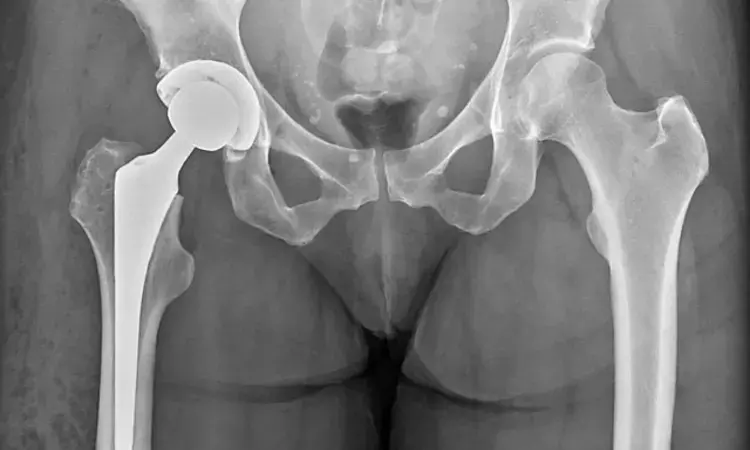
Hip X-rays helpful for osteoporosis screening in total hip arthroplasty patients

Germany: A recent study published in Scientific Reports has revealed the usefulness of plain hip X-rays to screen patients for osteoporosis before they undergo total hip arthroplasty (THA).
"An easily accessible screening tool for osteoporosis or osteopenia using plain hip radiographs is of great importance for orthopaedic hip surgeons to avoid potential surgical risks while performing THA due to osteoporosis and osteopenia," the researchers noted.
Previous studies have shown the underuse of dual-energy x-ray absorptiometry (DEXA) in patients before THA, despite being the gold standard for osteoporosis assessment. However, several studies have suggested that indices on plain hip X-rays may be reliable screening tools in Asian ethnicities or females.
Given the lack of knowledge about male patients and Caucasian ethnicities, Sebastian Rohe, the University Hospital Jena in Eisenberg, Germany, and colleagues aimed to evaluate plane hip radiographic indices as a screening tool for osteoporosis and osteopenia in Caucasian female and male patients before undergoing THA.
They investigated the correlation between indices on standardized preoperative X-rays of the hip and the proximal femoral neck T-score, measured by DEXA, in a Caucasian population with a high number of male patients.
The research team retrospectively analyzed results from 216 elderly female and male Caucasian patients treated with unilateral THA for primary osteoarthritis and who underwent preoperative DEXA scans and standard x-rays of the hip within one year. They correlated femoral neck DEXA T-scores with four indices on hip X-rays: canal calcar ratio (CCR), the canal flare index (CFI), and canal bone ratio (CBR) 7 cm and 10 cm below the lesser trochanter. A total of 216 patients (49.5% male) were included.
The study led to the following findings:
- CBR-7 and -10 were highly correlated with femoral neck T-score in males (Pearson’s correlation CBR-7 r = − 0.60, CBR-10 r = − 0.55) and females (r = − 0.74, r = − 0.77).
- CBR-7 and -10 also showed good diagnostic accuracy for osteoporosis in the ROC analysis in males (CBR-7: AUC = 0.75, threshold = 0.51; CBR-10: 0.63; 0.50) and females (CBR-7: AUC = 0.87, threshold = 0.55; CBR-10: 0.90; 0.54).
"Indices such as the CBR 7 or 10 cm below the lesser trochanter on plain hip radiographs are a good screening tool for osteopenia and osteoporosis on plain hip radiographs and can be used to initiate further diagnostics like the gold standard DXA," the researchers wrote. "They differ between male and female patients."
"In the future, the calculated values could be used by AI planning algorithms to recommend additional preoperative DXA to individualize the surgical procedure," they concluded.
Reference:
Rohe, S., Böhle, S., Matziolis, G., Jacob, B., & Brodt, S. (2023). Plain radiographic indices are reliable indicators for quantitative bone mineral density in male and female patients before total hip arthroplasty. Scientific Reports, 13(1), 1-8. https://doi.org/10.1038/s41598-023-47247-w
Dr Kamal Kant Kohli-MBBS, DTCD- a chest specialist with more than 30 years of practice and a flair for writing clinical articles, Dr Kamal Kant Kohli joined Medical Dialogues as a Chief Editor of Medical News. Besides writing articles, as an editor, he proofreads and verifies all the medical content published on Medical Dialogues including those coming from journals, studies,medical conferences,guidelines etc. Email: drkohli@medicaldialogues.in. Contact no. 011-43720751


