Modified 20% anatomical axis method demonstrates the highest reliability for posterior tibial slope measurement on radiographs: study
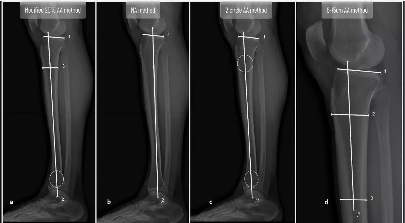


Several methods are described for the measurement of the posterior tibial slope (PTS), with no clear consensus on which method is the most reproducible and precise for measuring the PTS. Shradha A. Kulkarni et al conducted a study to identify the most reliable method for measuring PTS that is easily reproducible and precise.
Patients who underwent unilateral medial opening wedge high tibial osteotomy (MOWHTO) for varus deformity were retrospectively analyzed using their preoperative and 6-month postoperative electronic optical scan (EOS) scans for measurement of PTS. Four commonly used methods to measure PTS were used for comparison of intraobserver and interobserver reproducibility: the modified 20% anatomical axis (AA) method, the mechanical axis (MA) estimation method, the 2-circle AA method and the 5–15 cm AA method on short lateral knee radiographs. Three methods used AA estimation whereas, one method used MA estimation for measuring PTS. Two observers performed two measurement sessions >1 week apart for each patient. The intraclass correlation coefficient (ICC) and 95% confidence intervals (95% CI) were used to measure the intraobserver and interobserver reproducibility and precision for each method. Cohen's d measure was used to ascertain the effect size.
Results of the study were:
• Among the 30 patients analyzed for this study, the median age of the patients included was 46 years with 21 males and 9 females.
• The modified 20% method was consistently found to be the most reproducible and precise for measurement of PTS among all methods on comparing ICC and 95% CIs.
• The other methods had comparable values of ICC and 95% CIs.
• The mean difference in measured PTS when using short or long knee radiographs with AA estimation methods was negligible.
• The measured PTS was found to be, on an average, 2◦ higher when using AA estimation methods than the MA estimation methods.
The authors concluded – “The modified 20% AA method was found to be the most reproducible and reliable method for measuring PTS. There was negligible difference in the measured PTS on long and short radiographs using AA estimation methods. The measured PTS was 2◦ higher when utilizing AA estimation methods than the MA estimation methods. Each technique has been shown to have satisfactory reliability for clinical use, and increased familiarity may improve reliability and precision.”
Level of evidence: Retrospective observational study (Level IV).
For further details on the article refer to:
Modified 20% anatomical axis method demonstrates the highest reliability for posterior tibial slope measurement on radiographs
Shradha A. Kulkarni et al
Journal of ISAKOS 20 (2026) 101186
http://dx.doi.org/10.2106/JBJS.OA.25.00162