- Home
- Medical news & Guidelines
- Anesthesiology
- Cardiology and CTVS
- Critical Care
- Dentistry
- Dermatology
- Diabetes and Endocrinology
- ENT
- Gastroenterology
- Medicine
- Nephrology
- Neurology
- Obstretics-Gynaecology
- Oncology
- Ophthalmology
- Orthopaedics
- Pediatrics-Neonatology
- Psychiatry
- Pulmonology
- Radiology
- Surgery
- Urology
- Laboratory Medicine
- Diet
- Nursing
- Paramedical
- Physiotherapy
- Health news
- Fact Check
- Bone Health Fact Check
- Brain Health Fact Check
- Cancer Related Fact Check
- Child Care Fact Check
- Dental and oral health fact check
- Diabetes and metabolic health fact check
- Diet and Nutrition Fact Check
- Eye and ENT Care Fact Check
- Fitness fact check
- Gut health fact check
- Heart health fact check
- Kidney health fact check
- Medical education fact check
- Men's health fact check
- Respiratory fact check
- Skin and hair care fact check
- Vaccine and Immunization fact check
- Women's health fact check
- AYUSH
- State News
- Andaman and Nicobar Islands
- Andhra Pradesh
- Arunachal Pradesh
- Assam
- Bihar
- Chandigarh
- Chattisgarh
- Dadra and Nagar Haveli
- Daman and Diu
- Delhi
- Goa
- Gujarat
- Haryana
- Himachal Pradesh
- Jammu & Kashmir
- Jharkhand
- Karnataka
- Kerala
- Ladakh
- Lakshadweep
- Madhya Pradesh
- Maharashtra
- Manipur
- Meghalaya
- Mizoram
- Nagaland
- Odisha
- Puducherry
- Punjab
- Rajasthan
- Sikkim
- Tamil Nadu
- Telangana
- Tripura
- Uttar Pradesh
- Uttrakhand
- West Bengal
- Medical Education
- Industry
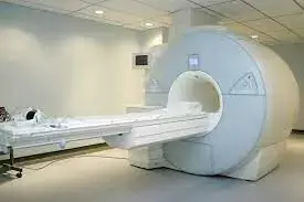
Novel Whole-body MRI may assess inflammatory arthritis in multiple joints in one examination: Study

Jane Freeston et al conducted a study to develop a novel whole-body MRI protocol capable of assessing inflammatory arthritis at an early stage in multiple joints in one examination.
Forty-six patients with inflammatory joint symptoms and 9 healthy volunteers underwent whole-body MR imaging on a 3.0 T MRI scanner in this prospective study. Image quality and pathology in each joint, bursae, entheses and tendons were scored by two of three radiologists and compared to clinical joint scores. Participants were divided into three groups based on diagnosis at 1-year follow-up (healthy volunteers, rheumatoid arthritis and all other types of arthritis). Radiology scores were compared between the three groups using a Kruskal-Wallis test. The clinical utility of radiology scoring was compared to clinical scoring using ROC analysis.
Key findings of the study:
• Fifty-five participants were included in this study comprising 18 patients with RA (10 cyclic citrullinated peptide (CCP) negative RA patients and 8 CCP positive RA patients), 28 patients with non-RA and 9 HC’s. The non RA group consisted of the following subtypes: 10 persistent undifferentiated arthritis (pUA), 8 resolved undifferentiated arthritis (rUA), 5 spondyloarthritis (SpA), 4 undifferentiated arthritis and 1 crystal arthritis.
• A protocol capable of whole-body MR imaging of the joints with an image acquisition time under 20 min was developed with excellent image quality.
• Synovitis scores were significantly higher in patients who were diagnosed with rheumatoid arthritis at 12 months (p < 0.05).
• Radiology scoring of bursitis showed statistically significant differences between each of the three groups—healthy control, rheumatoid arthritis and non-rheumatoid arthritis (p < 0.05).
• There was no statistically significant difference in ROC analysis between MRI and clinical scores.
The authors concluded –
“In summary, this whole-body multi-joint MRI technique is feasible to use in a clinical setting and produces good quality images. It has the potential to differentiate between RA, non-RA and HC at early presentation. It is potentially useful in identifying disease “load”, including sub-clinical disease and extra-articular inflammation. The technique is transferrable to other MRI scanners and to other patient cohorts where assessment of global joint-based disease is desirable.”
Further reading:
Whole body MRI for the investigation of joint involvement in inflammatory arthritis
Jane Freeston et al
Skeletal Radiology (2024) 53:935–945
https://doi.org/10.1007/s00256-023-04515-0
MBBS, Dip. Ortho, DNB ortho, MNAMS
Dr Supreeth D R (MBBS, Dip. Ortho, DNB ortho, MNAMS) is a practicing orthopedician with interest in medical research and publishing articles. He completed MBBS from mysore medical college, dip ortho from Trivandrum medical college and sec. DNB from Manipal Hospital, Bengaluru. He has expirence of 7years in the field of orthopedics. He has presented scientific papers & posters in various state, national and international conferences. His interest in writing articles lead the way to join medical dialogues. He can be contacted at editorial@medicaldialogues.in.


