- Home
- Medical news & Guidelines
- Anesthesiology
- Cardiology and CTVS
- Critical Care
- Dentistry
- Dermatology
- Diabetes and Endocrinology
- ENT
- Gastroenterology
- Medicine
- Nephrology
- Neurology
- Obstretics-Gynaecology
- Oncology
- Ophthalmology
- Orthopaedics
- Pediatrics-Neonatology
- Psychiatry
- Pulmonology
- Radiology
- Surgery
- Urology
- Laboratory Medicine
- Diet
- Nursing
- Paramedical
- Physiotherapy
- Health news
- Fact Check
- Bone Health Fact Check
- Brain Health Fact Check
- Cancer Related Fact Check
- Child Care Fact Check
- Dental and oral health fact check
- Diabetes and metabolic health fact check
- Diet and Nutrition Fact Check
- Eye and ENT Care Fact Check
- Fitness fact check
- Gut health fact check
- Heart health fact check
- Kidney health fact check
- Medical education fact check
- Men's health fact check
- Respiratory fact check
- Skin and hair care fact check
- Vaccine and Immunization fact check
- Women's health fact check
- AYUSH
- State News
- Andaman and Nicobar Islands
- Andhra Pradesh
- Arunachal Pradesh
- Assam
- Bihar
- Chandigarh
- Chattisgarh
- Dadra and Nagar Haveli
- Daman and Diu
- Delhi
- Goa
- Gujarat
- Haryana
- Himachal Pradesh
- Jammu & Kashmir
- Jharkhand
- Karnataka
- Kerala
- Ladakh
- Lakshadweep
- Madhya Pradesh
- Maharashtra
- Manipur
- Meghalaya
- Mizoram
- Nagaland
- Odisha
- Puducherry
- Punjab
- Rajasthan
- Sikkim
- Tamil Nadu
- Telangana
- Tripura
- Uttar Pradesh
- Uttrakhand
- West Bengal
- Medical Education
- Industry
Imageless Robotic Knee Arthroplasty: Novel surgical technique

Durham, NC: Robotic-assisted total knee arthroplasty is increasing in prevalence and has been shown to enable improved accuracy in implant positioning in total knee arthroplasty (TKA). Robotic assisted TKA can be categorized into image-guided and imageless techniques.
In image guided robotic TKA systems, preoperative imaging, most frequently computed tomography, is used to map bony anatomical landmarks to preoperatively obtained image to plan bone resections and implant sizing and positioning. Imageless robotic-assisted TKA does not require preoperative advanced imaging and intraoperatively maps bony anatomy to guide bone resection and implant sizing and placement.
Surgical Technique:
Room Setup and Patient Positioning
The room is setup so that the camera and monitor are opposite to the surgeon. The limb is prepped and draped in a sterile manner and placed in a knee positioner.
Exposure
A longitudinal incision is then made from the medial aspect of the tibial tubercle to the top of the patella with the knee in flexion, and a standard medial parapatellar arthrotomy is performed.
The incision is slightly longer proximally to accommodate femoral pin placement inside the wound.
In imageless robotic-assisted knee arthroplasty, the surgeon should identify and excise any prominent spurs or osteophytes of the femur, tibia, and patella prior to mapping the bony surfaces.
Thorough osteophyte excision facilitates proper surface mapping of the tibia and femur so that accurate preoperative bone morphology is obtained to optimally plan for final implant size and placement.
This is a key difference from image-guided systems where osteophytes and bone prominences should not be removed prior to registration since the preoperative template is based on them.
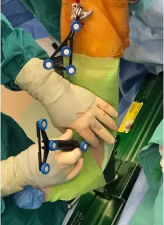
Next, the tibial and femoral tracking pins are placed. Tracker arrays and clamps are applied and tightened to the femur and tibia tracking pins. Tracker position should be confirmed so that the position of the camera cart and tracker arrays allow for full, uninterrupted visibility throughout the registration and cutting processes. At defined stages during the procedure, a point probe is put in contact with the clamp that attaches to the previously placed pins in the femur and tibia to ensure that the tracker arrays have not moved throughout the surgery.
Registration and Surface Mapping
After the hip, femur knee and tibia centers are collected, the knee is fully extended and flexed, with no varus or valgus stress applied. This determines neutral preoperative mechanical align ment, pre-operative extension, flexion, and kinematic axis.
A point probe is used to first outline and then fill the distal femur & proximal tibia.
Planning
The first step in planning is determining the size and position of the tibia and femur components. The computer will size and position the components based on registration data, which can then be modified based on surgeon preference.
In the gap assessment stage. Stressed ROM can be applied by placing a Z-retractor in the medial and lateral compartment for valgus and varus stress, respectively. Stressed ROM data is processed to produce a gap assessment graph that demonstrates the gap or overlap between the tibia and femur components throughout the range of motion in both the medial and lateral compartments.
Femoral and tibial cuts
All colored areas represent parts of the femur and tibia that need to be resected. Milling is performed from anterior to posterior down to the appropriate depth of the femur and tibia.
The tibial tray size is confirmed manually, and a tibial and femoral trial and trial liner are placed.
Guard comes off the burr, allowing for independent surgeon-driven maneuver (not computer software supported) to prepare the PS box.
Implant Trialing and Postop Assessment
The knee is again taken through both nonstressed and stressed ROM and a graph is plotted to allow the surgeon to compare postop stressed ROM from the initial planned stressed ROM.
Once satisfied with trial implants and postop gap assessment, trials are removed, bony surfaces are irrigated, and standard cementing technique can be used to implant final components.
The authors opined that - imageless robotic-assisted arthroplasty offers a reproducible, accurate, and more convenient approach to robotic-assisted TKA, with early studies reporting excellent short-term clinical outcomes and survivorship. With their high level of implant accuracy, they anticipate that these systems will translate to excellent medium and long-term clinical outcomes and survivorship, though more studies are warranted in this area.
Further reading:
Imageless Robotic Knee Arthroplasty
Mark Wu, Lefko Charalambous, Colin Penrose, Elshaday Belay, and Thorsten M. Seyler
Operative Techniques in Orthopaedics
Volume 31, Issue 4, December 2021, 100906
https://doi.org/10.1016/j.oto.2021.100906
MBBS, Dip. Ortho, DNB ortho, MNAMS
Dr Supreeth D R (MBBS, Dip. Ortho, DNB ortho, MNAMS) is a practicing orthopedician with interest in medical research and publishing articles. He completed MBBS from mysore medical college, dip ortho from Trivandrum medical college and sec. DNB from Manipal Hospital, Bengaluru. He has expirence of 7years in the field of orthopedics. He has presented scientific papers & posters in various state, national and international conferences. His interest in writing articles lead the way to join medical dialogues. He can be contacted at editorial@medicaldialogues.in.


