- Home
- Medical news & Guidelines
- Anesthesiology
- Cardiology and CTVS
- Critical Care
- Dentistry
- Dermatology
- Diabetes and Endocrinology
- ENT
- Gastroenterology
- Medicine
- Nephrology
- Neurology
- Obstretics-Gynaecology
- Oncology
- Ophthalmology
- Orthopaedics
- Pediatrics-Neonatology
- Psychiatry
- Pulmonology
- Radiology
- Surgery
- Urology
- Laboratory Medicine
- Diet
- Nursing
- Paramedical
- Physiotherapy
- Health news
- Fact Check
- Bone Health Fact Check
- Brain Health Fact Check
- Cancer Related Fact Check
- Child Care Fact Check
- Dental and oral health fact check
- Diabetes and metabolic health fact check
- Diet and Nutrition Fact Check
- Eye and ENT Care Fact Check
- Fitness fact check
- Gut health fact check
- Heart health fact check
- Kidney health fact check
- Medical education fact check
- Men's health fact check
- Respiratory fact check
- Skin and hair care fact check
- Vaccine and Immunization fact check
- Women's health fact check
- AYUSH
- State News
- Andaman and Nicobar Islands
- Andhra Pradesh
- Arunachal Pradesh
- Assam
- Bihar
- Chandigarh
- Chattisgarh
- Dadra and Nagar Haveli
- Daman and Diu
- Delhi
- Goa
- Gujarat
- Haryana
- Himachal Pradesh
- Jammu & Kashmir
- Jharkhand
- Karnataka
- Kerala
- Ladakh
- Lakshadweep
- Madhya Pradesh
- Maharashtra
- Manipur
- Meghalaya
- Mizoram
- Nagaland
- Odisha
- Puducherry
- Punjab
- Rajasthan
- Sikkim
- Tamil Nadu
- Telangana
- Tripura
- Uttar Pradesh
- Uttrakhand
- West Bengal
- Medical Education
- Industry
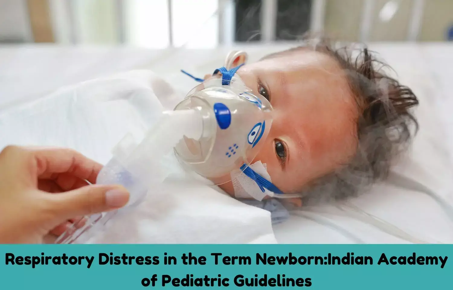
Respiratory Distress in the Term Newborn: IAP Guidelines

Respiratory distress (RD) in newborn is characterized by increased work of breathing (WOB) in the form of tachypnea, grunting, chest retractions, and often associated with reduced air entry and cyanosis.
The Indian Academy of Pediatrics (IAP) has released Standard Treatment Guidelines 2022 for Respiratory Distress in the Term Newborn. The lead author for these guidelines on Respiratory Distress in the Term Newborn is Dr. Srinivas Murki along with co-author Dr. Umamaheswari B and Dr. Rameshwor Yengkhom. The guidelines come Under the Auspices of the IAP Action Plan 2022, and the members of the IAP Standard Treatment Guidelines Committee include Chairperson Remesh Kumar R, IAP Coordinator Vineet Saxena, National Coordinators SS Kamath, Vinod H Ratageri, Member Secretaries Krishna Mohan R, Vishnu Mohan PT and Members Santanu Deb, Surender Singh Bisht, Prashant Kariya, Narmada Ashok, Pawan Kalyan.
Following are the major recommendations of guidelines:
Assessment of Severity:
Downes and Silverman Anderson Score (SAS) on the clinical evaluation, oxygen saturation (SpO2 ) and fraction of inspired oxygen (FiO2 ) requirement, oxygen saturation index (OSI), alveolar-arterial diffusion gradient of oxygen (A-aDO2 ), oxygenation index (OI), and arterial blood gas parameters are useful in the assessment of severity of RD in a term infant. There are various clinical scoring systems for assessing the severity of RD objectively, out of which Downes scoring (Tables 1) and Silverman Anderson (Tables 2) scoring systems are widely used. Downes scoring system is used for term neonates whereas SAS score is often used in preterm neonates. A total score of 0 suggests no distress, score of 1–4 mild RD, score of 5–7 moderate RD, and score of >7 severe distress or impending respiratory failure.
| TABLE 1: Downes score. | |||||
| Score | Respiratory rate | Cyanosis | Air entry | Grunt | Retraction |
| 0 | <60 breaths/minute | Nil | Normal | None | Nil |
| 1 | 60–80 breaths/minute | In room air | Mild decrease | Audible with stethoscope | Mild |
| 2 | >80 breaths/minute or apnea | In >40% oxygen | Marked decrease | Audible without stethoscope | Moderate- to-severe |
| TABLE 2: Silverman Anderson score (SAS). | |||||
| Score | Upper chest* | Lower chest# | Xiphoid retractions | Nares dilatation | Grunting |
| 0 | Synchronized | No retractions | None | None | None |
| 1 | Lag on inspiration | Just visible | Just visible | Minimal | Heard with stethoscope |
| 2 | Seesaw | Marked | Marked | Marked | Heard without stethoscope |
Saturation index = (MAP × FiO2)/SpO2
A-aDO2 = (700 × FiO2) – (PaCO2 + PaO2) or
= (760* – Water vapor pressure × FiO2) – (PaCO2/0.8#) – PaO2
PF ratio = PaO2/FiO2
Oxygenation index = (MAP × FiO2)/PaO2
*760 denotes the atmospheric pressure at sea level
#0.8 denotes respiratory quotient
(MAP: mean airway pressure; FiO2: fraction of inspired oxygen; PaO2 and PaCO2: calculated from arterial blood gas; SpO2: saturation from pulse oximeter)
Alveolar–Arterial Diffusion Gradient of Oxygen:
A-aDO2 is the difference between amount of oxygen in alveoli and the amount of oxygen dissolved in plasma (arterial oxygenation).
A-aDO2 values could reach up to 200–400 in severe RD syndrome, persistent pulmonary hypertension (PPHN) and severe meconium aspiration syndrome (MAS).
PF Ratio (PaO2 /FiO2 ):
This is one of the measures used in ventilated neonates. Ratio of <300 mm Hg indicates abnormal gas exchange.
Oxygenation Index:
This is commonly used in neonates to assess the severity and to guide on the timing of intervention.
OI value of <15 suggest mild HRF; 15–25 suggests moderate HRF; values >25 suggest severe HRF, and the need for inhaled nitric oxide (iNO) therapy.
A persistent value above 40 is an indication for extracorporeal membrane oxygenation (ECMO).
Blood Gas Analysis:
Arterial or capillary blood gas analysis helps in assessing the severity of RD and guiding the management.
Normal range of blood gas values in neonates are:
• pH 7.35–7.45, PaCO2 35–45 mm Hg, PaO2 45–80 mm Hg, bicarbonate 20–24 mEq/L, and base deficit 3–7 mEq/L.
Etiology:
Causes of RD in term neonates are depicted in Table 3.
| TABLE 3: Causes of respiratory distress. | |
| Common causes | Uncommon causes |
|
and birth injury |
| TABLE 4: Etiology according to the onset of respiratory distress. | |
| Onset | Etiology |
| <6 hours of life |
þ Early-onset pneumonia/sepsis þ Meconium aspiration syndrome (MAS) þ Perinatal asphyxia þ Congenital diaphragmatic hernia (CDH) |
| >6–12 hours of life |
þ Pneumonia
þ Hypothermia and hypoglycemia þ Inborn error of metabolism þ Congenital pulmonary airway malformation (CPAM) |
| TABLE 5: Etiology based on history. | |
| History | Presentation |
| History of maternal diabetes | RDS, TTN, MAS, and asphyxia |
| History of maternal fever/PPROM/chorioamnionitis | Sepsis and pneumonia |
| Fetal distress/CTG abnormality | Asphyxia |
| Elective cesarean section without labor | TTN |
| Consanguinity/previous sibling death | Inborn errors of metabolism (IEM) |
| Antenatal scan abnormality:
| Tracheoesophageal fistula, CDH, and CPAM Pulmonary hypoplasia CDH, CPAM, pleural effusion/hydrops, and congenital heart disease |
| TABLE 6: Chest X-ray and ultrasound findings in various conditions. | ||
| Condition | Chest X-ray | Ultrasound lung |
| TTN | Sun burst appearance; fluid in minor fissure | Thickened pleural lines, B lines, double lung point |
| MAS | Hyperinflation with bilateral patchy lung opacities | Disappearance of A lines, scattered B lines |
| RDS | Reticulogranular opacities/ground glass appearance | B lines, white lungs |
| Pneumonia | Asymmetrical parenchymal infilterates | Nonspecific changes |
| Pneumothorax | Collapsed lung border with air in pleural space with mediastinal shift | Absence of sliding sign; Bar code sign |
| TABLE 7: Differences between CHD and pulmonary disease. | ||
| Pulmonary disease | Cyanotic heart disease | |
| Onset of respiratory distress | Since birth or within 6 hours of life | Usually after 24 hours, when the ductus arteriosus closes |
| History | Risk factors such as maternal fever, prolonged rupture of membranes, and meconium-stained amniotic fluid could be elicited | Family history of congenital heart disease may be seen |
| Antenatal scans | Could detect congenital malformations such as CDH, CPAM, tracheoesophageal fistula | Structural heart conditions could have been detected in antenatal scans |
| Respiratory distress | Usually moderate to severe distress associated with chest retractions | Silent tachypnea in cardiac conditions with reduced pulmonary flow; mild- to-moderate distress in conditions with increased pulmonary blood flow |
| Other signs | Scaphoid abdomen and hyperinflated chest in CDH; copious secretion in TEF; septic shock can be present in pneumonia; barrel-shaped chest in MAS; labile saturations; and hypoxia during handling in PPHN | Cyanosis, murmur, signs of cardiac failure (gallop rhythm, and hepatomegaly), prominent precordial pulsations, single second heart sound, and feeble femoral pulses |
| Pulse oximetry screening | Occasionally positive (false positive in PPHN and certain respiratory conditions) | Positive with greater accuracy |
| Arterial blood gas | Hypoxia (PaO2 low) Hypercapnia (PaCO2 high) | Hypoxia (PaO2 low) Hypocarbia or normocarbia (PaCO2 normal or low) |
| Hyperoxia test* | PaO2 > 150 mm Hg | PaO2 < 150 mm Hg |
| Chest X-ray | No cardiomegaly Patchy consolidation in pneumonia; bilateral patchy infiltrates with hyperinflation-MAS; prominent bronchovascular marking and fluid in minor fissure TTN; normal lungs in PPHN; ground glass appearance in RDS | Egg on side appearance in TGA; normal or small heart with pulmonary edema in obstructive TAPVC; box- shaped heart in Ebstein anomaly |
| Echocardiography | Structurally normal heart; could show features of PPHN | Confirms the diagnosis |
- General Therapy
- Surfactants
- Antibiotics
- Inotropes
- Pulmonary Vasodilators
(A-aDO2 : alveolar-arterial diffusion gradient of oxygen; CPAP: continuous positive airway pressure; CXR: chest X-ray; FiO2 : fraction of inspired oxygen; HFNC: high-flow nasal cannula; NIV: noninvasive ventilation; OI: oxygenation index; PCV: packed cell volume; RD: respiratory distress; SpO2 : oxygen saturation; WOB: work of breathing)
- Cloherty JP, Eichenwald EC, Hansen AR, Stark AR. Manual of Neonatal Care, 7th edition. Netherlands: Wolters Kluwer; 2012.
- Edwards MO, Kotecha SJ, Kotecha S. Respiratory distress of the term newborn infant. Paediatr Respir Rev. 2013;14:29-37.
- Mathai SS, Raju U, Kanitkar M. Management of respiratory distress in the newborn. Med J Armed Forces India. 2007;63(3):269-72.
- Pramanik AK, Rangaswamy N, Gates T. Neonatal respiratory distress: a practical approach to its diagnosis and management. Pediatr Clin North Am. 2015;62(2):453-69.
- Reuter S, Moser C, Baack M. Respiratory distress in the newborn. Pediatr Rev. 2014;35;417-28.
- Taeusch HW, Ballard RA, Avery ME, Gleason CA. Avery's Diseases of the Newborn, 10th edition. Amsterdam, Netherlands: Elsevier; 2018.
The guidelines can be accessed on the official site of IAP: https://iapindia.org/standard-treatment-guidelines/
I have done my Bachelor of pharmacy from United Institute of Pharmacy and currently pursuing pharmaceutical MBA from Jamia hamdard. I worked as an intern at the position of content creator in Medical Dialogue and am highly obliged to the company for giving me this wonderful opportunity.
Dr Kamal Kant Kohli-MBBS, DTCD- a chest specialist with more than 30 years of practice and a flair for writing clinical articles, Dr Kamal Kant Kohli joined Medical Dialogues as a Chief Editor of Medical News. Besides writing articles, as an editor, he proofreads and verifies all the medical content published on Medical Dialogues including those coming from journals, studies,medical conferences,guidelines etc. Email: drkohli@medicaldialogues.in. Contact no. 011-43720751


