- Home
- Medical news & Guidelines
- Anesthesiology
- Cardiology and CTVS
- Critical Care
- Dentistry
- Dermatology
- Diabetes and Endocrinology
- ENT
- Gastroenterology
- Medicine
- Nephrology
- Neurology
- Obstretics-Gynaecology
- Oncology
- Ophthalmology
- Orthopaedics
- Pediatrics-Neonatology
- Psychiatry
- Pulmonology
- Radiology
- Surgery
- Urology
- Laboratory Medicine
- Diet
- Nursing
- Paramedical
- Physiotherapy
- Health news
- Fact Check
- Bone Health Fact Check
- Brain Health Fact Check
- Cancer Related Fact Check
- Child Care Fact Check
- Dental and oral health fact check
- Diabetes and metabolic health fact check
- Diet and Nutrition Fact Check
- Eye and ENT Care Fact Check
- Fitness fact check
- Gut health fact check
- Heart health fact check
- Kidney health fact check
- Medical education fact check
- Men's health fact check
- Respiratory fact check
- Skin and hair care fact check
- Vaccine and Immunization fact check
- Women's health fact check
- AYUSH
- State News
- Andaman and Nicobar Islands
- Andhra Pradesh
- Arunachal Pradesh
- Assam
- Bihar
- Chandigarh
- Chattisgarh
- Dadra and Nagar Haveli
- Daman and Diu
- Delhi
- Goa
- Gujarat
- Haryana
- Himachal Pradesh
- Jammu & Kashmir
- Jharkhand
- Karnataka
- Kerala
- Ladakh
- Lakshadweep
- Madhya Pradesh
- Maharashtra
- Manipur
- Meghalaya
- Mizoram
- Nagaland
- Odisha
- Puducherry
- Punjab
- Rajasthan
- Sikkim
- Tamil Nadu
- Telangana
- Tripura
- Uttar Pradesh
- Uttrakhand
- West Bengal
- Medical Education
- Industry
Subpleural nodules and septal thickening diagnostic of pulmonary TB with pleural effusion on CT chest: Study

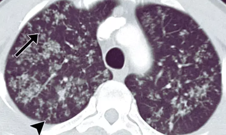
South Korea: A new study published in CHEST Journal by Min Kyung Jung and a team of researchers suggests that computed tomography (CT) findings of subpleural micronodules and interlobular septal thickening could aid in differentiating tuberculous (TB) pleural effusion from non-tuberculous empyema.
The research, published in a reputable medical journal, provides valuable insights into the CT characteristics of TB-related pleural effusion and their association with specific imaging features.
The study aimed to investigate the correlation between the frequency of subpleural micronodules, interlobular septal thickening, and the presence of pleural effusion in patients with pulmonary TB. The researchers retrospectively analyzed pulmonary TB patients' CT findings, including micronodule distribution, significant opacity, cavitation, tree-in-buds, bronchovascular bundle thickening, interlobular septal thickening, lymphadenopathy, and pleural effusion.
- Of the 338 patients diagnosed with pulmonary TB who underwent CT scans, 60 were excluded due to co-existing pulmonary diseases.
- The remaining patients were divided into two groups based on the presence or absence of pleural effusion.
- The analysis revealed significant differences in the frequency of subpleural nodules and interlobular septal thickening between the two groups.
- Subpleural nodules were more prevalent in patients with TB pleural effusion, with a frequency of 69% compared to 14% in those without effusion.
- Interlobular septal thickening was also more commonly observed in the pleural effusion group (81%) compared to the non-effusion group (64%).
- On the other hand, tree-in-buds were less frequently seen in patients with TB pleural effusion (29%) than those without effusion (48%).
The findings suggest that subpleural nodules and interlobular septal thickening on CT scans can provide essential clues in diagnosing TB pleural effusion. These CT features may indicate TB involvement of the lymphatics in the peripheral interstitium, which could be associated with the development of pleural effusion.
This research contributes to our understanding of the CT characteristics of TB-related pleural effusion and may have implications for clinical practice. By identifying specific imaging features, healthcare professionals can more accurately differentiate TB pleural effusion from non-TB empyema, improving diagnostic accuracy and time management.
Further studies are warranted to validate these findings and explore the potential utility of CT findings in diagnosing and managing TB-related pleural effusion. By leveraging imaging technology, healthcare providers can enhance their ability to detect and differentiate various pleural pathologies, ultimately improving patient outcomes and optimizing treatment strategies for TB-related conditions.
Reference:
Jung, M. K., Lee, S. Y., Min, E. J., & Ko, J. M. (2023). CT differences of pulmonary tuberculosis according to presence of pleural effusion. Chest. https://doi.org/10.1016/j.chest.2023.06.043
Dr Kamal Kant Kohli-MBBS, DTCD- a chest specialist with more than 30 years of practice and a flair for writing clinical articles, Dr Kamal Kant Kohli joined Medical Dialogues as a Chief Editor of Medical News. Besides writing articles, as an editor, he proofreads and verifies all the medical content published on Medical Dialogues including those coming from journals, studies,medical conferences,guidelines etc. Email: drkohli@medicaldialogues.in. Contact no. 011-43720751


