- Home
- Medical news & Guidelines
- Anesthesiology
- Cardiology and CTVS
- Critical Care
- Dentistry
- Dermatology
- Diabetes and Endocrinology
- ENT
- Gastroenterology
- Medicine
- Nephrology
- Neurology
- Obstretics-Gynaecology
- Oncology
- Ophthalmology
- Orthopaedics
- Pediatrics-Neonatology
- Psychiatry
- Pulmonology
- Radiology
- Surgery
- Urology
- Laboratory Medicine
- Diet
- Nursing
- Paramedical
- Physiotherapy
- Health news
- Fact Check
- Bone Health Fact Check
- Brain Health Fact Check
- Cancer Related Fact Check
- Child Care Fact Check
- Dental and oral health fact check
- Diabetes and metabolic health fact check
- Diet and Nutrition Fact Check
- Eye and ENT Care Fact Check
- Fitness fact check
- Gut health fact check
- Heart health fact check
- Kidney health fact check
- Medical education fact check
- Men's health fact check
- Respiratory fact check
- Skin and hair care fact check
- Vaccine and Immunization fact check
- Women's health fact check
- AYUSH
- State News
- Andaman and Nicobar Islands
- Andhra Pradesh
- Arunachal Pradesh
- Assam
- Bihar
- Chandigarh
- Chattisgarh
- Dadra and Nagar Haveli
- Daman and Diu
- Delhi
- Goa
- Gujarat
- Haryana
- Himachal Pradesh
- Jammu & Kashmir
- Jharkhand
- Karnataka
- Kerala
- Ladakh
- Lakshadweep
- Madhya Pradesh
- Maharashtra
- Manipur
- Meghalaya
- Mizoram
- Nagaland
- Odisha
- Puducherry
- Punjab
- Rajasthan
- Sikkim
- Tamil Nadu
- Telangana
- Tripura
- Uttar Pradesh
- Uttrakhand
- West Bengal
- Medical Education
- Industry
Low-dose three-phase brain perfusion imaging and AI-based parameter map generation:Study

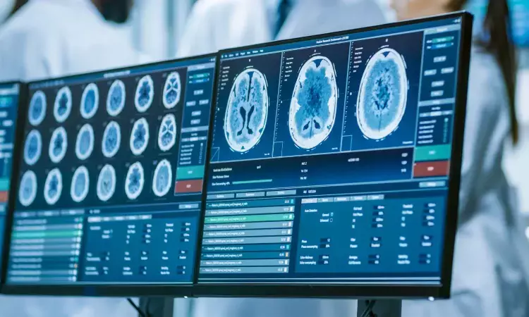
Computed Tomography Perfusion (CTP) is a critical tool for rapidly evaluating brain blood flow in suspected stroke patients, guiding time-sensitive treatment decisions. However, standard CTP requires continuously scanning the brain over 40-60 seconds, capturing numerous time points. This results in high cumulative radiation doses, poses risks for patients with kidney problems due to the contrast agent load, and is sensitive to patient movement, leading to complex processing and potential failure. While reducing the number of scans seems logical, randomly skipping time points often misses crucial peaks in blood flow, severely underestimating key parameters. Thus, how to optimize scanning protocols to reduce CTP radiation dose and address the complexity of current functional imaging processes are urgent issues to address.
Now, team from the First Affiliated Hospital of Jinan University and Southern Medical University, have developed an innovative CTP scanning protocol and a deep learning model that can generate the vital blood flow maps needed to assess stroke patients. This work has proofed the proposed low radiation dose imaging program can slash radiation exposure by over 80% compared to current methods. This innovation promised to make stroke diagnosis safer and more accessible, particularly for vulnerable patients.
Addressing Limitations in Conventional CTP
Despite its clinical value, traditional CTP is associated with significant drawbacks. These include:
- High Radiation Dose: Conventional protocols can reach cumulative doses around 5260 mGy·cm, notably higher than CTA (~3222 mGy·cm).
- Motion Sensitivity: Repeated scans across timepoints make the technique vulnerable to patient motion, requiring sophisticated registration algorithms.
- Workflow Complexity: The image processing burden and risk of failure hinder routine use in clinical settings.
Previous strategies for reducing radiation via temporal subsampling risk omitting critical arterial enhancement peaks, underestimating hemodynamic parameters. While multiphase CTA (mCTA) has shown promise in capturing arterial and venous phases, it requires large contrast volumes (~80 mL), posing risks for patients with impaired renal function, and lacks quantitative perfusion data.
A Three-Phase CTP with Deep Learning Enhancement
Inspired by the temporal structure of mCTA, the team introduced a three-phase CTP protocol that drastically reduces temporal sampling while preserving essential perfusion information. A generative adversarial network (GAN)-based model was developed to directly synthesize perfusion parameter maps from only three timepoints.
In internal validation datasets, the model-produced maps showed high structural and perceptual fidelity compared to ground truth, demonstrating its capability to reconstruct key perfusion features. Further experiments explored how variations in the selected three-phase combinations affected performance. Even with ±2-second deviations from the ideal timepoints, the model maintained high predictive accuracy, although performance dropped with deviations beyond 4 seconds. These findings support both the practical feasibility of the protocol and the robustness of the model.
Reference:
Cuidie Zeng, Xiaoling Wu, Fusheng Ouyang, Baoliang Guo, Xiao Zhang, Jianghua Ma, Dong Zeng, Bin Zhang. Perfusion Parameter Map Generation from 3 Phases of Computed Tomography Perfusion in Stroke Using Generative Adversarial Networks. Research. 2025;8:0689.DOI:10.34133/research.0689.
Dr Kamal Kant Kohli-MBBS, DTCD- a chest specialist with more than 30 years of practice and a flair for writing clinical articles, Dr Kamal Kant Kohli joined Medical Dialogues as a Chief Editor of Medical News. Besides writing articles, as an editor, he proofreads and verifies all the medical content published on Medical Dialogues including those coming from journals, studies,medical conferences,guidelines etc. Email: drkohli@medicaldialogues.in. Contact no. 011-43720751


