- Home
- Medical news & Guidelines
- Anesthesiology
- Cardiology and CTVS
- Critical Care
- Dentistry
- Dermatology
- Diabetes and Endocrinology
- ENT
- Gastroenterology
- Medicine
- Nephrology
- Neurology
- Obstretics-Gynaecology
- Oncology
- Ophthalmology
- Orthopaedics
- Pediatrics-Neonatology
- Psychiatry
- Pulmonology
- Radiology
- Surgery
- Urology
- Laboratory Medicine
- Diet
- Nursing
- Paramedical
- Physiotherapy
- Health news
- Fact Check
- Bone Health Fact Check
- Brain Health Fact Check
- Cancer Related Fact Check
- Child Care Fact Check
- Dental and oral health fact check
- Diabetes and metabolic health fact check
- Diet and Nutrition Fact Check
- Eye and ENT Care Fact Check
- Fitness fact check
- Gut health fact check
- Heart health fact check
- Kidney health fact check
- Medical education fact check
- Men's health fact check
- Respiratory fact check
- Skin and hair care fact check
- Vaccine and Immunization fact check
- Women's health fact check
- AYUSH
- State News
- Andaman and Nicobar Islands
- Andhra Pradesh
- Arunachal Pradesh
- Assam
- Bihar
- Chandigarh
- Chattisgarh
- Dadra and Nagar Haveli
- Daman and Diu
- Delhi
- Goa
- Gujarat
- Haryana
- Himachal Pradesh
- Jammu & Kashmir
- Jharkhand
- Karnataka
- Kerala
- Ladakh
- Lakshadweep
- Madhya Pradesh
- Maharashtra
- Manipur
- Meghalaya
- Mizoram
- Nagaland
- Odisha
- Puducherry
- Punjab
- Rajasthan
- Sikkim
- Tamil Nadu
- Telangana
- Tripura
- Uttar Pradesh
- Uttrakhand
- West Bengal
- Medical Education
- Industry
Ultra-low-dose of CT in 3D printing models good enough to diagnose wrist fractures

The introduction of CT in the early 1970s provided orthopaedic surgeons with a method of imaging that could be used to characterise a fracture more completely than by conventional radiography alone.
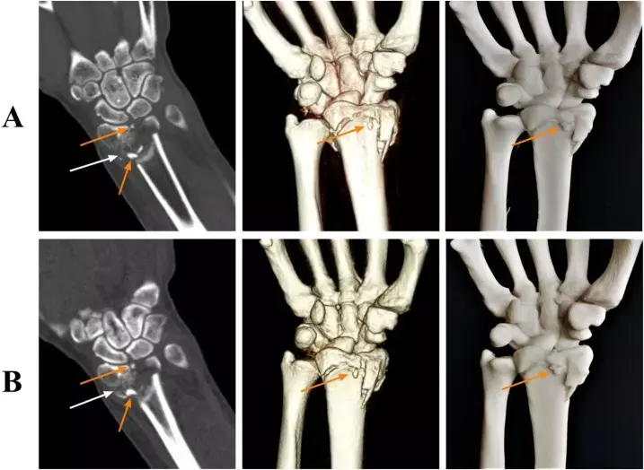
Today, CT scanning is used routinely in the evaluation of specific fractures. A research team of Guangdong Hospital of Traditional Chinese Medicine, China recently found that an ultralow-dose CT protocol yielded 97% lower radiation dose and enabled the production of 3D-printed models that were deemed equivalent to those manufactured from standard-dose images for distal radial fractures. The research findings were published in the European Journal of Radiology on December 29, 2020.
CT has several advantages, including the ability to characterise a fracture in greater detail, to identify an occult fracture, and to diagnose incomplete union, all of which have practical implications for treatment of the individual patient. For these reasons, the number of CT scans performed annually has increased dramatically. Bone injuries can be treated using tissue engineering scaffolds, but the conventional constructs have a big challenge in supplying requirements of native tissue, i.e., bioactivity potential, mechanical stability, controllable biodegradability, and proper cellular interaction. In this regard, 3D printing technology with the possibility of controlling the internal microstructure and geometry of synthesized matrixes was introduced as a promising approach for bone defect regeneration. However, the effects of low-dose CT techniques on 3D printing have been unknown. Therefore, the researchers conducted a study to explore the effect of ultra-low-dose computed tomography (CT) on three-dimensional (3D) printing models and the diagnosis of wrist fractures.
They enrolled a total of 76 consecutive patients with clinical suspicion of distal radial fractures (DRFs). All individuals had undergone 320-row detector CT and researchers randomly divided them into Group A (n=38) that received unenhanced CT scans at standard-dose (120 kV and 100 mA) and Group B (n=38) that received ultralow-dose (80 kV and 10 mA) protocols on an Aquilion One CT scanner. For objective image quality assessment, they measured the noise, CT number, signal-to-noise ratio (SNR), and contrast-to-noise ratio (CNR). Two orthopaedic surgeons blinded to the scan parameters evaluated the clarity of the 3D printing model and fracture line using a 3-point scale. Researchers also calculated the mean radiation dose and compared diagnostic performances for the fractures between the two groups.
They found that the effective radiation dose was significantly reduced by 97.1 % in Group B when compared with Group A. They also found the Quantitative objective image quality parameters (e.g., CNR, SNR, and CT numbers) were higher in the standard-dose group. However, they found no difference in subjective scoring of the 3D printing model. Although the fracture line score was higher in Group A they noted a consistent diagnostic performance in both the groups. They observed no significant differences in the sensitivity, specificity or accuracy between standard-dose group and ultra-low-dose group.
The authors concluded, "The ultra-low-dose protocol effectively reduced the radiation dose by 97.1 %, while maintaining the image quality for the diagnosis of DRFs. Therefore, this protocol can meet the needs of 3D printing models for preoperative assessments".
For further information:
Medical Dialogues Bureau consists of a team of passionate medical/scientific writers, led by doctors and healthcare researchers. Our team efforts to bring you updated and timely news about the important happenings of the medical and healthcare sector. Our editorial team can be reached at editorial@medicaldialogues.in.
Dr Kamal Kant Kohli-MBBS, DTCD- a chest specialist with more than 30 years of practice and a flair for writing clinical articles, Dr Kamal Kant Kohli joined Medical Dialogues as a Chief Editor of Medical News. Besides writing articles, as an editor, he proofreads and verifies all the medical content published on Medical Dialogues including those coming from journals, studies,medical conferences,guidelines etc. Email: drkohli@medicaldialogues.in. Contact no. 011-43720751


