- Home
- Medical news & Guidelines
- Anesthesiology
- Cardiology and CTVS
- Critical Care
- Dentistry
- Dermatology
- Diabetes and Endocrinology
- ENT
- Gastroenterology
- Medicine
- Nephrology
- Neurology
- Obstretics-Gynaecology
- Oncology
- Ophthalmology
- Orthopaedics
- Pediatrics-Neonatology
- Psychiatry
- Pulmonology
- Radiology
- Surgery
- Urology
- Laboratory Medicine
- Diet
- Nursing
- Paramedical
- Physiotherapy
- Health news
- Fact Check
- Bone Health Fact Check
- Brain Health Fact Check
- Cancer Related Fact Check
- Child Care Fact Check
- Dental and oral health fact check
- Diabetes and metabolic health fact check
- Diet and Nutrition Fact Check
- Eye and ENT Care Fact Check
- Fitness fact check
- Gut health fact check
- Heart health fact check
- Kidney health fact check
- Medical education fact check
- Men's health fact check
- Respiratory fact check
- Skin and hair care fact check
- Vaccine and Immunization fact check
- Women's health fact check
- AYUSH
- State News
- Andaman and Nicobar Islands
- Andhra Pradesh
- Arunachal Pradesh
- Assam
- Bihar
- Chandigarh
- Chattisgarh
- Dadra and Nagar Haveli
- Daman and Diu
- Delhi
- Goa
- Gujarat
- Haryana
- Himachal Pradesh
- Jammu & Kashmir
- Jharkhand
- Karnataka
- Kerala
- Ladakh
- Lakshadweep
- Madhya Pradesh
- Maharashtra
- Manipur
- Meghalaya
- Mizoram
- Nagaland
- Odisha
- Puducherry
- Punjab
- Rajasthan
- Sikkim
- Tamil Nadu
- Telangana
- Tripura
- Uttar Pradesh
- Uttrakhand
- West Bengal
- Medical Education
- Industry
Pre-operative CT evaluation essential for planning proper pedicle screw placement in AIS patients

Pre-operative CT evaluation is essential for planning proper pedicle screw placement in AIS patients suggests a new study published in the BMC Surgery.
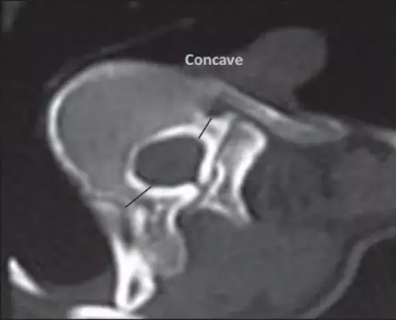
Screw insertion during scoliosis surgery uses free-hand pedicle screw insertion methods. However, there is a wide variation in pedicle shapes, sizes, and morphometry, especially in scoliosis patients. CT scan pedicle measurements in main thoracic Lenke type 1 adolescent idiopathic scoliosis can help visualize this diversity. This study aimed to highlight the features of pedicle morphometry on the concave and convex sides, including pedicle diameter (width in axial and height in the sagittal plane), the depth to the anterior cortex, and Watanabe Pedicle classification in patients with main thoracic apex adolescent idiopathic scoliosis.
This study was a cross-sectional observational study of Adolescent Idiopathic Scoliosis (AIS) patients whose apex in the main thoracic patient underwent deformity correction procedures. We used a three-dimensional CT scan to evaluate pedicle morphometry on the apex vertebrae, three consecutive vertebrae above and below the apex.
Results
A total of 6 patients with apex main thoracic AIS with 84 pedicles consisting of 42 pedicles from each concave and convex curve were analyzed.
• All of the samples were female, with the mean age at the procedure being 21.2 ± 5.56.
• The mean cobb angle was 62° ± 23°, with the main apex between VT8-VT10.
• The size of the pedicle was bigger from upper to lower vertebrae.
• The mean pedicle depth, pedicle width, and pedicle height for the concave side were 36.06 ± 4.31 mm, 3.91 ± 0.66 mm, and 9.16 ± 1.52 mm, respectively.
• Meanwhile, the convex side is 37.52 ± 1.84 mm, 5.20 ± 0.55 mm, and 11.05 ± 0.70 mm, respectively.
• They found a significant difference between the concave and convex sides for the pedicle width and height.
• The concave and convex sides were mainly classified as type C (38%) and type A (50%) Watanabe pedicle.
Pedicle width and pedicle height are significantly different between the concave and the convex side with convex side has better Watanabe pedicle classification. Pre-operative CT evaluation is essential for planning proper pedicle screw placement in AIS patients. f
Reference:
Sakti, Y.M., Lanodiyu, Z.A., Ichsantyaridha, M. et al. Pedicle morphometry analysis of main thoracic apex adolescent idiopathic scoliosis. BMC Surg 23, 34 (2023).https://doi.org/10.1186/s12893-022-01877-5
Keywords:
Pedicle morphometry, Morphology, CT scan, Adolescent idiopathic scoliosis, Main thoracic apex, Sakti, Y.M., Lanodiyu, Z.A., Ichsantyaridha, M, BMC surgery
Dr. Shravani Dali has completed her BDS from Pravara institute of medical sciences, loni. Following which she extensively worked in the healthcare sector for 2+ years. She has been actively involved in writing blogs in field of health and wellness. Currently she is pursuing her Masters of public health-health administration from Tata institute of social sciences. She can be contacted at editorial@medicaldialogues.in.
Dr Kamal Kant Kohli-MBBS, DTCD- a chest specialist with more than 30 years of practice and a flair for writing clinical articles, Dr Kamal Kant Kohli joined Medical Dialogues as a Chief Editor of Medical News. Besides writing articles, as an editor, he proofreads and verifies all the medical content published on Medical Dialogues including those coming from journals, studies,medical conferences,guidelines etc. Email: drkohli@medicaldialogues.in. Contact no. 011-43720751


