- Home
- Medical news & Guidelines
- Anesthesiology
- Cardiology and CTVS
- Critical Care
- Dentistry
- Dermatology
- Diabetes and Endocrinology
- ENT
- Gastroenterology
- Medicine
- Nephrology
- Neurology
- Obstretics-Gynaecology
- Oncology
- Ophthalmology
- Orthopaedics
- Pediatrics-Neonatology
- Psychiatry
- Pulmonology
- Radiology
- Surgery
- Urology
- Laboratory Medicine
- Diet
- Nursing
- Paramedical
- Physiotherapy
- Health news
- Fact Check
- Bone Health Fact Check
- Brain Health Fact Check
- Cancer Related Fact Check
- Child Care Fact Check
- Dental and oral health fact check
- Diabetes and metabolic health fact check
- Diet and Nutrition Fact Check
- Eye and ENT Care Fact Check
- Fitness fact check
- Gut health fact check
- Heart health fact check
- Kidney health fact check
- Medical education fact check
- Men's health fact check
- Respiratory fact check
- Skin and hair care fact check
- Vaccine and Immunization fact check
- Women's health fact check
- AYUSH
- State News
- Andaman and Nicobar Islands
- Andhra Pradesh
- Arunachal Pradesh
- Assam
- Bihar
- Chandigarh
- Chattisgarh
- Dadra and Nagar Haveli
- Daman and Diu
- Delhi
- Goa
- Gujarat
- Haryana
- Himachal Pradesh
- Jammu & Kashmir
- Jharkhand
- Karnataka
- Kerala
- Ladakh
- Lakshadweep
- Madhya Pradesh
- Maharashtra
- Manipur
- Meghalaya
- Mizoram
- Nagaland
- Odisha
- Puducherry
- Punjab
- Rajasthan
- Sikkim
- Tamil Nadu
- Telangana
- Tripura
- Uttar Pradesh
- Uttrakhand
- West Bengal
- Medical Education
- Industry
Ten Futuristic Applications of a Basic Ultrasound
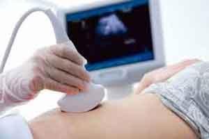
Here are a few futuristic expansions in the use of this very flexible cross-sectional modality, scans possible in all three axis, which will enhance the importance of this tool in clinical practice.
Musculoskeletal Ultrasound
Ultrasound of bones and joints or Musculoskeletal Ultrasounds have been slowly gaining strong popularity in the country, with its strong importance in detection of fractures and diseases in patients where MRI and CT cannot be used or is difficult, particularly with small children and older patients. It has variety of uses including diagnosing infections of bone, joints, and soft tissue, foreign body detection, tumors, soft tissue injuries and variety of other situations where MRI will be like killing an ant with a cannon ball, Also presence of pacemakers, stents, implants, etc are relative contraindications for performing MRI.
Dermatology/ Skin Ultrasound
Ultrasounds can be used for detection and diagnosis of many skin diseases. These range from detection and monitoring of benign as well as malignant skin tumours , vascular lesions, skin inflammatory diseases, and other nail lesions. Of-Late, Derma ultrasounds are also being used in cosmetology, being used to assess skin’s response to treatments such as laser, mesotherapy, photodynamic therapy, etc
[caption id="attachment_10050" align="aligncenter" width="420"]
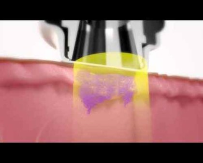 Image Source: article.wn.com[/caption]
Finger/ Hand/ Nail Ultrasounds
Modalities of CT-scan and even MRIs face a strong limitation when it comes to diagnosis of problems in hands and fingers. MRI of the fingers and hands can only be done, if you have the part- specific coil and not all MRI machines are equipped with the same. Hence it becomes difficult sometimes or impossible to use an MRI for hands and fingers for accurate diagnosis. In these situations ultrasounds can be helpful in detection of a number of problems including fractures, detection of foreign bodies, tumours and masses, as well as diseases and infection of tendons. It is also being widely used for procedures inclduding guidance of injection, aspiration or biopsy.
Off lately, ultrasounds of nails have also become popular for detection and diagnosis of many diseases including tumours of the nail, infections of the bone (osteomyelitis) occult fractures of the phallanges, Epidermoids or inclusion dermoids ( eg in seamstresses), Viability of the tissues through colour doppler scans for the interdigital and nail bed vascularity. etc
[caption id="attachment_10049" align="aligncenter" width="420"]
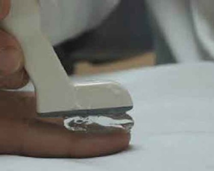 Image Source: www.researchgate.net[/caption]
Facial Ultrasounds.
Ultrasound now is increasingly being used for diagnosis of various facial bones. Ocular ultrasounds are coming up for detection of various diseases including eye ball perforation, retrobulbar hematoma / tumours, retinal detachment, lens subluxation, vitreous hemorrhage, and intraocular foreign.
[caption id="attachment_10082" align="aligncenter" width="266"]
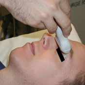 Image Source: sonoguide.com[/caption]
Similarly we have ultrasounds of the TM joints for osteoarthritis secondary to teeth grinding (bruxism) dislocations and subluxations, occult fractures neck mandible , and also cosmetic redesigning of hypoplastic mandible.
Dental Ultrasounds
While facial ultrasound is one sphere, ultrasound is also gaining importance in the sphere of dentistry as a painless modality to detect tooth lesions and defects, oral ultrasound with water contrast study for evaluation of mucosal lesion and gingival thickness, dental implant locations, as well as even periodontal bony disorders as also foreign body localization
[caption id="attachment_10052" align="aligncenter" width="420"]
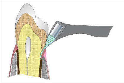 Image SOurce: spinoff.nasa.gov[/caption]
Cranial/Brain Ultrasounds
Ultrasounds in the form of intra-op Brain ultrasounds as well as trans-cranial dopplers are increasingly being used to evaluate the brain tissue and the flow of blood to the brain. While brain ultrasounds are predominantly used in infants ( whose bone has not developed ) through the fontenelle, TCD ultrasounds evaluate the direction and velocity of the blood flow in the major cerebral arteries of the brain through the thin temporal bone for conditions such as Giant cell arteritis
[caption id="attachment_10084" align="aligncenter" width="380"]
 Image Source: http://www.armobgyn.com[/caption]
Lung Ultrasounds
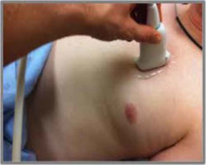
In the past decade, use of Ultrasound for diagnosis of diseases of the lung has seen a sharp rise specially in the emergency rooms / casualties Apart from the traditional use of ultrasound to detect pleural effusions and masses, ultrasounds are now increasing being used to in the evaluation of many different acute and chronic conditions, from cardiogenic pulmonary edema to acute lung injury, from pneumothorax to pneumonia, from interstitial lung disease to pulmonary infarctions and contusions (MS)
[caption id="attachment_10048" align="aligncenter" width="420"]
 www.medspecindiana.com[/caption]
Emergency Ultrasounds
From traditionally being placed in the corners of the diagnostic departments of the hospitals, ultrasound machines are slowly finding their way to the front board of ERs/ Emergencies and Casualties, with the modality coming in handy for any and many kinds of emergencies including trauma, injuries.
[caption id="attachment_10057" align="aligncenter" width="275"]
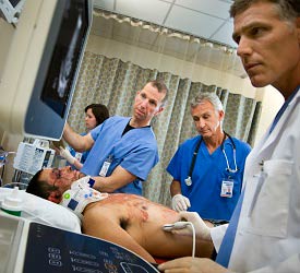 Image Source: Healthcare.phillips.com[/caption]
Although this is now a norm in the west, Indian hospitals are slowly bringing in the use of this modality for quick and painless detection in the face of emergencies by techniques such as e-FAST .
ICU Based Ultrasounds
[caption id="attachment_10055" align="aligncenter" width="290"]
 Image Source: research gate.net[/caption]
The importance of ultrasound in an ICU setting can never be undermined. With recent developments the scope of ultrasound has expanded in the sphere of intensive care. Think of a difficult intubation or even a bloodless tracheostomy, ultrasound is regularly being used in both diagnostic as well as therapeutic procedures. Oesophageal intubations are a nightmare in emergency settings and ultrasound proves to be a handy tool in its detection.
Pediatric Ultrasounds
[caption id="attachment_10087" align="aligncenter" width="400"]
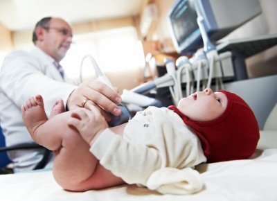 Image Source: http://www.ultrasoundtechniciancenter.org[/caption]
Ultrasound is quite a useful modality when it comes to small children. Modalities of CT and MRI become restricted when it comes to children, who get scared and claustrophobic with the machines and have to be given sedatives for the use of the modality. Ultrasound is Man –send since CT in growing skeletons is a relative contraindication. ALARA is our mantra today in evaluation of pediatric patients keeping radiation as low as possible and MRI with the absolute need for sedation becomes difficult to use. Ultrasound is the modality of choice for preliminary scanning to localize the pathology and initiate treatment be it musculoskeletal, lung abdomen, pseudo-paralysis.
Radiology
Dr Nidhi Bhatnagar MBBS, MD, PGDHM, is a radiologist presently working as Principal Consultant, Radiology Department, Max Superspeciality Centre, Panchsheel, New Delhi. Earlier she was the Head of the Department, Radiology, Chanan Devi Hospital C1, Janak Puri, New Delhi. She was the Faculty- Guest speaker at Delhi Orthopedic Association Conference, Dec. 2011, on “Case presentations- Role of USG in diagnosis of chronic osteomyelitis”. She was the Professor, Universidad Catolica San Antonio Murcia, Spain. Musculoskeletal Ultrasound, Faculty and Guide of National Board of Examinations, Radio-diagnosis, Editor at Musculoskeletal Imaging, Apollo Medicine Journal, Reviewer, IJRI (Indian Journal of radiology and Imaging) , MSK US. She was the Editorial Manager of the the online submission and peer review tracking system for American Journal of Roentgenology (AJR). She was the Woman Achiever 2020 in the Field of Radiology, Felicitated by IRIA and ICRI. She was the Founder Member and General Secretary of Muskuloskeletal Ultrasound Society (MUS), and the Founder member Musculoskeletal Imaging Foundation, (MIF) Ganga Hospital, Coimbatore. Intellectual rights 2016, Govt of India , Created a web-based software with a patent in the name of Dr Nidhi Bhatnagar, the purpose of filling F-forms on–line, data audit, saving the information and for the purpose of data mining. She was the Past Joint Secretary of Delhi state Chapter, Indian Radiological and Imaging Association (IRIA) 2014, Ex-member to the Committee of State Appropriate Authority for Inspection and Monitoring .


