
Research finds AI Can Classify TEE Views and Cardiac Function During Surgery
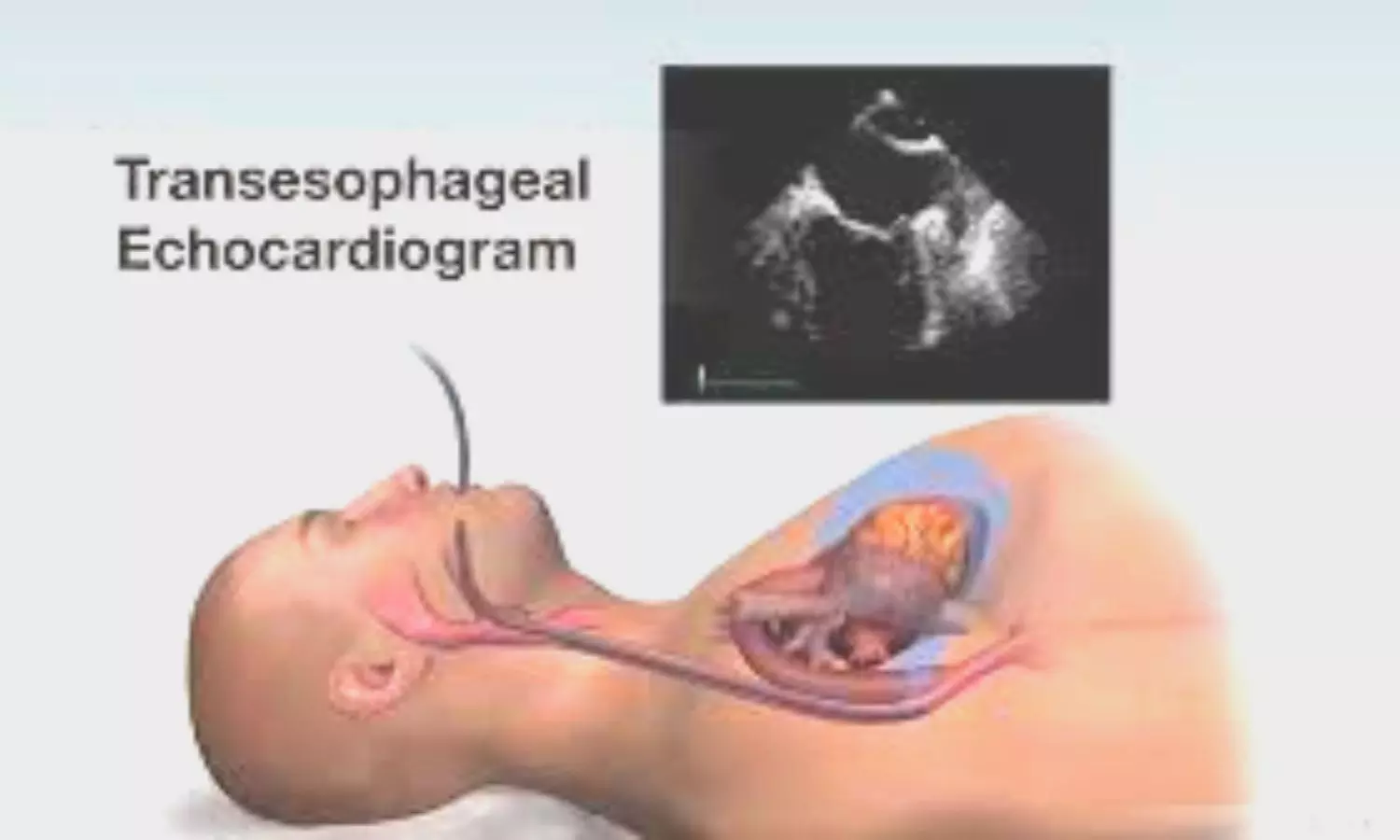



AI assistant in the cardiac OR that instantly interprets your transesophageal echocardiography (TEE) images—view classification, ejection fraction, right ventricular function, and even tricuspid regurgitation—before you can even call for a consult. This scenario is no longer science fiction. A new proof-of-concept study in the Journal of Cardiothoracic and Vascular Anesthesia demonstrates that deep learning can automate comprehensive intraoperative TEE analysis with promising accuracy.
Study Design: A Deep Dive Into TEE Automation
Researchers conducted a cross-sectional study using more than 700,000 TEE video clips from 6,900 cardiac surgery cases at two hospitals. The automated framework was trained to handle three core tasks:
View classification: Correctly identifying 26 standard TEE views
Biventricular function assessment: Determining left ventricular ejection fraction (LVEF) and right ventricular systolic function (RVSF)
Tricuspid regurgitation (TR) grading: Differentiating levels of TR severity
Expert-annotated datasets and state-of-the-art neural networks (including convolutional and vision transformer models) powered the system, which was tested against hundreds of held-out studies.
Key Findings: High Accuracy Across Core Diagnostic Tasks
View classification reached 86% accuracy, outperforming human expert agreement (74%).
The model distinguished normal vs. abnormal LVEF with AUROC up to 0.95 (for severe dysfunction cutoffs).
Right ventricular dysfunction was identified with AUROC of 0.92 for moderate or greater impairment.
Tricuspid regurgitation grading was more challenging but achieved AUROC of 0.79 for detecting moderate or worse cases.
Continuous LVEF prediction showed strong correlation (Pearson r = 0.79) with a mean absolute error of 7.8%.
Technical Note:
AUROC (Area Under the Receiver Operating Characteristic curve) quantifies model diagnostic accuracy; values close to 1.0 are considered excellent.
Convolutional neural networks and vision transformers are deep learning methods that analyze spatial and temporal image features.
Why This Matters: Potential for Real-Time, Reliable Decision Support
For anesthesiologists and cardiac surgeons, intraoperative TEE is critical—but interpretation is operator-dependent and time-consuming. This study’s automated TEE framework promises:
Standardized view classification: Reducing subjectivity in image interpretation
Fast, reproducible function assessment: Enabling more confident, data-driven intraoperative decisions
Scalability: Framework can be retrained and adapted to additional cardiac parameters in future
While automated TTE (transthoracic echo) solutions are gaining traction, this is one of the first large studies to rigorously tackle the complexity of intraoperative TEE.
Limitations and Next Steps
The framework was internally validated but not yet tested in external hospital systems. Ground-truth labels relied on clinical reports rather than quantitative measurements, and other intraoperative findings (e.g., diastolic function, prosthetic valve assessment) were not included. Further work is needed to validate the model in different settings, improve interpretability, and integrate with clinical workflows.
Conclusion
This proof-of-concept study demonstrates that deep learning can automate complex TEE interpretation tasks—including view classification and biventricular function assessment—at a level comparable to expert clinicians. As AI tools continue to evolve, these innovations may soon support real-time, standardized diagnostic guidance during cardiac surgery.
KEY POINTS
Automated AI framework classified intraoperative TEE views with 86% accuracy—outperforming human expert agreement.
Model accurately predicted left and right ventricular function and tricuspid regurgitation severity using deep learning.
System analyzed over 700,000 TEE clips from nearly 7,000 cardiac surgery cases.
AI-driven TEE interpretation could improve intraoperative decision-making and workflow efficiency.
External validation and broader clinical deployment are future priorities for this technology.
Citation:
Chan T, Goldfinger S, Grasfield RH, Eswar V, Barreto D, Augoustides JG, Yan V, Shah RM, Pouch AM, MacKay EJ. Proof-of-Concept Automated Framework for Intraoperative Transesophageal Echocardiography: View Classification and Biventricular Function Assessment. Journal of Cardiothoracic and Vascular Anesthesia. 2026;40:1339–1348. DOI: https://doi.org/10.1053/j.jvca.2026.01.036