- Home
- Medical news & Guidelines
- Anesthesiology
- Cardiology and CTVS
- Critical Care
- Dentistry
- Dermatology
- Diabetes and Endocrinology
- ENT
- Gastroenterology
- Medicine
- Nephrology
- Neurology
- Obstretics-Gynaecology
- Oncology
- Ophthalmology
- Orthopaedics
- Pediatrics-Neonatology
- Psychiatry
- Pulmonology
- Radiology
- Surgery
- Urology
- Laboratory Medicine
- Diet
- Nursing
- Paramedical
- Physiotherapy
- Health news
- Fact Check
- Bone Health Fact Check
- Brain Health Fact Check
- Cancer Related Fact Check
- Child Care Fact Check
- Dental and oral health fact check
- Diabetes and metabolic health fact check
- Diet and Nutrition Fact Check
- Eye and ENT Care Fact Check
- Fitness fact check
- Gut health fact check
- Heart health fact check
- Kidney health fact check
- Medical education fact check
- Men's health fact check
- Respiratory fact check
- Skin and hair care fact check
- Vaccine and Immunization fact check
- Women's health fact check
- AYUSH
- State News
- Andaman and Nicobar Islands
- Andhra Pradesh
- Arunachal Pradesh
- Assam
- Bihar
- Chandigarh
- Chattisgarh
- Dadra and Nagar Haveli
- Daman and Diu
- Delhi
- Goa
- Gujarat
- Haryana
- Himachal Pradesh
- Jammu & Kashmir
- Jharkhand
- Karnataka
- Kerala
- Ladakh
- Lakshadweep
- Madhya Pradesh
- Maharashtra
- Manipur
- Meghalaya
- Mizoram
- Nagaland
- Odisha
- Puducherry
- Punjab
- Rajasthan
- Sikkim
- Tamil Nadu
- Telangana
- Tripura
- Uttar Pradesh
- Uttrakhand
- West Bengal
- Medical Education
- Industry
From pulp to cementum: 3D visualization of soft and hard dental tissues using different ex vivo nano-CT contrast-enhancement techniques

In a new study, researchers found that decalcification followed by Lugol's iodine treatment significantly improved soft tissue contrast, particularly for pulp visualisation. In contrast, PTA without decalcification provided better contrast for dentine and allowed clear visualisation of attached soft tissues like the periodontal ligament and predentine. These results help guide the selection of imaging protocols tailored to specific dental tissue analyses.
A study was done to determine the effect of two contrast-enhancement strategies in nano-computed tomography (nano-CT) imaging on the contrast-to-noise ratio (CNR) of various dental tissues, including pulp, dentine and cementum, with the goal of enhancing the visibility of dental soft tissues to a level not yet reported in laboratory nano-CT imaging. Ten sound human third molars underwent decalcification and subsequent treatment with Lugol's iodine (n = 5, Group 1) or phosphotungstic acid (PTA) treatment without prior decalcification (n = 5, Group 2) for contrast enhancement.
Imaging was performed using the laboratory nano-CT system Skyscan 2211 and the synchrotron radiation for medical physics (SYRMEP) beamline. CNRs were measured for pulpal tissue, dentine and cementum and nano-CT images were compared with classical histology, scanning electron microscopy (SEM) and energy-dispersive X-ray spectroscopy (EDXS). Results: Group 1 significantly enhanced the contrast of pulp tissue, resulting in a 168.2% increase due to decalcification and an additional 148.7% increase after Lugol's iodine treatment. Dentine exhibited higher contrast in Group 2, whereas cementum showed similar contrast across both groups.
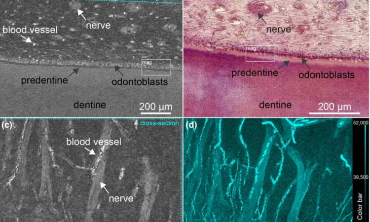
Laboratory nano-CT enabled the visualization of detailed soft tissue structures, including nerves, blood vessels and odontoblasts within the pulp, but cementocytes remained invisible. Decalcification followed by Lugol's iodine treatment was superior for enhancing soft tissue contrast, especially for pulp visualization. PTA without decalcification yielded better contrast for dentine and facilitated the visualization of attached soft tissues, such as periodontal ligament and predentine.
These findings provide insights into selecting the most appropriate protocol to optimize nano-CT imaging for specific dental tissue analyses, including the pulp.
Reference:
Hildebrand, T., Haugen, H.J., Romandini, M., Plotino, G. & Nogueira, L.P. (2025) From pulp to cementum: 3D visualization of soft and hard dental tissues using different ex vivo nano-CT contrast-enhancement techniques. International Endodontic Journal, 00, 1–15. Available from: https://doi.org/10.1111/iej.14260
Dr. Shravani Dali has completed her BDS from Pravara institute of medical sciences, loni. Following which she extensively worked in the healthcare sector for 2+ years. She has been actively involved in writing blogs in field of health and wellness. Currently she is pursuing her Masters of public health-health administration from Tata institute of social sciences. She can be contacted at editorial@medicaldialogues.in.
Dr Kamal Kant Kohli-MBBS, DTCD- a chest specialist with more than 30 years of practice and a flair for writing clinical articles, Dr Kamal Kant Kohli joined Medical Dialogues as a Chief Editor of Medical News. Besides writing articles, as an editor, he proofreads and verifies all the medical content published on Medical Dialogues including those coming from journals, studies,medical conferences,guidelines etc. Email: drkohli@medicaldialogues.in. Contact no. 011-43720751


