- Home
- Medical news & Guidelines
- Anesthesiology
- Cardiology and CTVS
- Critical Care
- Dentistry
- Dermatology
- Diabetes and Endocrinology
- ENT
- Gastroenterology
- Medicine
- Nephrology
- Neurology
- Obstretics-Gynaecology
- Oncology
- Ophthalmology
- Orthopaedics
- Pediatrics-Neonatology
- Psychiatry
- Pulmonology
- Radiology
- Surgery
- Urology
- Laboratory Medicine
- Diet
- Nursing
- Paramedical
- Physiotherapy
- Health news
- Fact Check
- Bone Health Fact Check
- Brain Health Fact Check
- Cancer Related Fact Check
- Child Care Fact Check
- Dental and oral health fact check
- Diabetes and metabolic health fact check
- Diet and Nutrition Fact Check
- Eye and ENT Care Fact Check
- Fitness fact check
- Gut health fact check
- Heart health fact check
- Kidney health fact check
- Medical education fact check
- Men's health fact check
- Respiratory fact check
- Skin and hair care fact check
- Vaccine and Immunization fact check
- Women's health fact check
- AYUSH
- State News
- Andaman and Nicobar Islands
- Andhra Pradesh
- Arunachal Pradesh
- Assam
- Bihar
- Chandigarh
- Chattisgarh
- Dadra and Nagar Haveli
- Daman and Diu
- Delhi
- Goa
- Gujarat
- Haryana
- Himachal Pradesh
- Jammu & Kashmir
- Jharkhand
- Karnataka
- Kerala
- Ladakh
- Lakshadweep
- Madhya Pradesh
- Maharashtra
- Manipur
- Meghalaya
- Mizoram
- Nagaland
- Odisha
- Puducherry
- Punjab
- Rajasthan
- Sikkim
- Tamil Nadu
- Telangana
- Tripura
- Uttar Pradesh
- Uttrakhand
- West Bengal
- Medical Education
- Industry
Dermoscopic features of acute cutaneous lupus erythematosus

Source- Behera B, Palit A, Sethy M, Nayak AK, Dash S, Ayyanar P. Dermoscopic features of acute cutaneous lupus erythematosus: A retrospective analysis from a tertiary care centre of East India. Australas J Dermatol. 2021 Aug;62(3):364-369. doi: 10.1111/ajd.13620. Epub 2021 May 25. PMID: 34033122.
Dermoscopic features of acute cutaneous lupus erythematosus
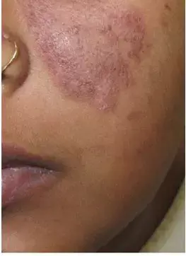
Acute cutaneous lupus erythematosus (ACLE) is a common clinical manifestation of active systemic lupus erythematosus (SLE). It can be localised or generalised. The localised form can be confused with rosacea, changes from chronic topical steroid use, demodicosis and seborrhoeic dermatitis. Similarly, generalised ACLE can be mistaken for atopic dermatitis, contact dermatitis, maculopapular drug rash, viral exanthem and rarely dermatomyositis. Dermoscopy can be a helpful tool to differentiate ACLE from other diseases. Recently the dermoscopic features of ACLE were reported in the Australasian Journal of Dermatology.
It was a retrospective study conducted at a tertiary care center of eastern India. All biopsy and direct immunofluorescence confirmed ACLE cases between May 2019 and May 2020 were included. The clinical and demographic data of the patients were retrieved from departmental database. Dermoscopic analysis was performed separately for localised and generalised ACLE.
A total of 21 patients (18 females and three males, aged range 4–52 years, with skin phototypes III, IV or V) were included in the analysis. Of the 21 patients, 17 had generalised ACLE and 9 had localised ACLE. The patients either had cutaneous lesions for the first time or had an exacerbation of the disease on presentation.
In both localised and generalised ACLE, the authors observed a multicomponent pattern comprising white scales, homogenous pinkish-white to reddish-white areas, brown dots/globules/peppering, keratotic follicular plugging and linear vessels with or without branching. The colour of keratotic follicular plugging was light to dark brown, except for yellow keratotic plugging in case of malar rash. There was a paucity of dotted vessels.
In ACLE, the dermoscopic examination reflects hyperkeratosis, keratotic plugging, dilated and/ or increased vessels as white homogenous area, keratotic plugging, linear vessels with or without branching, and/ or red/ pink homogenous area, respectively.
The presence of brown dots, globules and peppering in the present study was due to the dermal melanin and melanophages (along with atrophic epidermis, a feature more marked in dark-skinned individual. In dark-skinned patients with ACLE, the presence of brown dots/globules/peppering, which results from interface dermatitis, can rule out dermatoses, such as atopic dermatitis, seborrhoeic dermatitis, sebopsoriasis, rosacea, changes from chronic topical steroid use and drug rash. Presence of follicular plugging can differentiate ACLE from dermatomyositis
In conclusion, this article describes the dermoscopic features of both localised and generalised ACLE in patients with dark skin. A multicomponent pattern comprising white scales, homogenous pinkish-white to reddish-white area, brown dots/globules/peppering, keratotic follicular plugging, linear vessels with or without branching may point to the diagnosis of ACLE.
Source- Behera B, Palit A, Sethy M, Nayak AK, Dash S, Ayyanar P. Dermoscopic features of acute cutaneous lupus erythematosus: A retrospective analysis from a tertiary care centre of East India. Australas J Dermatol. 2021 Aug;62(3):364-369. doi: 10.1111/ajd.13620. Epub 2021 May 25. PMID: 34033122.
MBBS
Dr Manoj Kumar Nayak has completed his M.B.B.S. from the prestigious institute Bangalore medical college and research institute, Bengaluru. He completed his M.D. Dermatology from AIIMS Rishikesh. He is actively involved in the field of dermatology with special interests in vitiligo, immunobullous disorders, psoriasis and procedural dermatology. His continued interest in academics and recent developments serves as an inspiration to work with medical dialogues.He can be contacted at editorial@medicaldialogues.in.
Dr Kamal Kant Kohli-MBBS, DTCD- a chest specialist with more than 30 years of practice and a flair for writing clinical articles, Dr Kamal Kant Kohli joined Medical Dialogues as a Chief Editor of Medical News. Besides writing articles, as an editor, he proofreads and verifies all the medical content published on Medical Dialogues including those coming from journals, studies,medical conferences,guidelines etc. Email: drkohli@medicaldialogues.in. Contact no. 011-43720751


