- Home
- Medical news & Guidelines
- Anesthesiology
- Cardiology and CTVS
- Critical Care
- Dentistry
- Dermatology
- Diabetes and Endocrinology
- ENT
- Gastroenterology
- Medicine
- Nephrology
- Neurology
- Obstretics-Gynaecology
- Oncology
- Ophthalmology
- Orthopaedics
- Pediatrics-Neonatology
- Psychiatry
- Pulmonology
- Radiology
- Surgery
- Urology
- Laboratory Medicine
- Diet
- Nursing
- Paramedical
- Physiotherapy
- Health news
- Fact Check
- Bone Health Fact Check
- Brain Health Fact Check
- Cancer Related Fact Check
- Child Care Fact Check
- Dental and oral health fact check
- Diabetes and metabolic health fact check
- Diet and Nutrition Fact Check
- Eye and ENT Care Fact Check
- Fitness fact check
- Gut health fact check
- Heart health fact check
- Kidney health fact check
- Medical education fact check
- Men's health fact check
- Respiratory fact check
- Skin and hair care fact check
- Vaccine and Immunization fact check
- Women's health fact check
- AYUSH
- State News
- Andaman and Nicobar Islands
- Andhra Pradesh
- Arunachal Pradesh
- Assam
- Bihar
- Chandigarh
- Chattisgarh
- Dadra and Nagar Haveli
- Daman and Diu
- Delhi
- Goa
- Gujarat
- Haryana
- Himachal Pradesh
- Jammu & Kashmir
- Jharkhand
- Karnataka
- Kerala
- Ladakh
- Lakshadweep
- Madhya Pradesh
- Maharashtra
- Manipur
- Meghalaya
- Mizoram
- Nagaland
- Odisha
- Puducherry
- Punjab
- Rajasthan
- Sikkim
- Tamil Nadu
- Telangana
- Tripura
- Uttar Pradesh
- Uttrakhand
- West Bengal
- Medical Education
- Industry
Dermoscopy can help diagnose AVH, obviating need for biopsy: Study

Source- Genedy R, Taha A. Acrokeratosis verruciformis of Hopf: dermoscopic and histopathological study of two siblings. Clin Exp Dermatol. 2021 Oct;46(7):1313-1314. doi: 10.1111/ced.14691. Epub 2021 May 10. PMID: 33866599.
Acrokeratosis verruciformis of Hopf: dermoscopic and histopathological study- Acrokeratosis verruciformis of Hopf (AVH) is an autosomal dominant genodermatosis. It is caused by a mutation in the ATP2A2 gene, located on chromosome 12q24. Both AVH and Darier disease are believed to be allelic disorders. Familial cases of AVH usually have early onset either at birth or childhood, while sporadic cases usually present later in life. Recently dermoscopy of AVH has been described in Clinical and Experimental Dermatology journal.
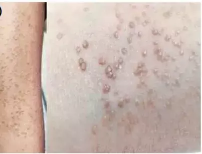
Two siblings, a 7-year-old boy and a 5-year-old girl born to nonconsanguineous parents presented with a 4-year history of multiple, asymptomatic, skin-coloured papules. The lesions were present mainly over the dorsal aspects of the forearms in the boy and ventral aspect of the forearm and the antecubital fossa in the girl.
Both patients had Fitzpatrick skin type IV. Rest of the cutaneous, mucosa, hair and nail examination were within normal limits. An excisional biopsy from a papule on each child showed a focal area of compact hyperkeratosis, papillomatosis with church spire appearance, hypergranulosis and acanthosis confirming the diagnosis of AVH.
Dermoscopic examination using polarized contact dermoscopy of the two patients revealed structureless skin coloured to yellowish areas with whitish dots and streaks. Some papules, especially the larger and less keratotic lesions, showed uniformly distributed dotted vessels. Few papules showed a cerebriform pattern too. Under nonpolarized contact dermoscopy, the papules featured irregular white homogeneous areas, a white network and a cobblestone appearance.
A recently published case by Rajan et al also showed central white homogeneous areas, a central white network, peripheral cobblestone appearance, multiple brown pigmented dots arranged radially in the periphery (sunray appearance) over acral papules and regularly arranged brown dots with background erythema in the nonlesional skin on polarized dermoscopy.2
AVH is a chronic disease characterized by flat-topped keratotic papules, typically observed on the dorsa of the hands and feet. The classic histopathological features include hyperkeratosis, papillomatosis with 'church spire' appearance, acanthosis and hypergranulosis. Clefts in the granular layer with several acantholytic keratinocytes have also been reported in AVH. So histopathology finally confirms the diagnosis.
To conclude dermoscopy can be helpful to diagnosis AVH and may help to eliminate the need for biopsy for diagnosis but robust studies on the disease are required to finally arrive at a conclusion.
Source-
1. Genedy R, Taha A. Acrokeratosis verruciformis of Hopf: dermoscopic and histopathological study of two siblings. Clin Exp Dermatol. 2021 Oct;46(7):1313-1314. doi: 10.1111/ced.14691. Epub 2021 May 10. PMID: 33866599.
2. Rajan MB, Bains A, Vedant D. Dermoscopy of acrokeratosis verruciformis of hopf. Indian Dermatol Online J 2021; 12: 374–5.
MBBS
Dr Manoj Kumar Nayak has completed his M.B.B.S. from the prestigious institute Bangalore medical college and research institute, Bengaluru. He completed his M.D. Dermatology from AIIMS Rishikesh. He is actively involved in the field of dermatology with special interests in vitiligo, immunobullous disorders, psoriasis and procedural dermatology. His continued interest in academics and recent developments serves as an inspiration to work with medical dialogues.He can be contacted at editorial@medicaldialogues.in.
Dr Kamal Kant Kohli-MBBS, DTCD- a chest specialist with more than 30 years of practice and a flair for writing clinical articles, Dr Kamal Kant Kohli joined Medical Dialogues as a Chief Editor of Medical News. Besides writing articles, as an editor, he proofreads and verifies all the medical content published on Medical Dialogues including those coming from journals, studies,medical conferences,guidelines etc. Email: drkohli@medicaldialogues.in. Contact no. 011-43720751


