- Home
- Medical news & Guidelines
- Anesthesiology
- Cardiology and CTVS
- Critical Care
- Dentistry
- Dermatology
- Diabetes and Endocrinology
- ENT
- Gastroenterology
- Medicine
- Nephrology
- Neurology
- Obstretics-Gynaecology
- Oncology
- Ophthalmology
- Orthopaedics
- Pediatrics-Neonatology
- Psychiatry
- Pulmonology
- Radiology
- Surgery
- Urology
- Laboratory Medicine
- Diet
- Nursing
- Paramedical
- Physiotherapy
- Health news
- Fact Check
- Bone Health Fact Check
- Brain Health Fact Check
- Cancer Related Fact Check
- Child Care Fact Check
- Dental and oral health fact check
- Diabetes and metabolic health fact check
- Diet and Nutrition Fact Check
- Eye and ENT Care Fact Check
- Fitness fact check
- Gut health fact check
- Heart health fact check
- Kidney health fact check
- Medical education fact check
- Men's health fact check
- Respiratory fact check
- Skin and hair care fact check
- Vaccine and Immunization fact check
- Women's health fact check
- AYUSH
- State News
- Andaman and Nicobar Islands
- Andhra Pradesh
- Arunachal Pradesh
- Assam
- Bihar
- Chandigarh
- Chattisgarh
- Dadra and Nagar Haveli
- Daman and Diu
- Delhi
- Goa
- Gujarat
- Haryana
- Himachal Pradesh
- Jammu & Kashmir
- Jharkhand
- Karnataka
- Kerala
- Ladakh
- Lakshadweep
- Madhya Pradesh
- Maharashtra
- Manipur
- Meghalaya
- Mizoram
- Nagaland
- Odisha
- Puducherry
- Punjab
- Rajasthan
- Sikkim
- Tamil Nadu
- Telangana
- Tripura
- Uttar Pradesh
- Uttrakhand
- West Bengal
- Medical Education
- Industry
Sarcoidosis of the ear, nose and throat: A review

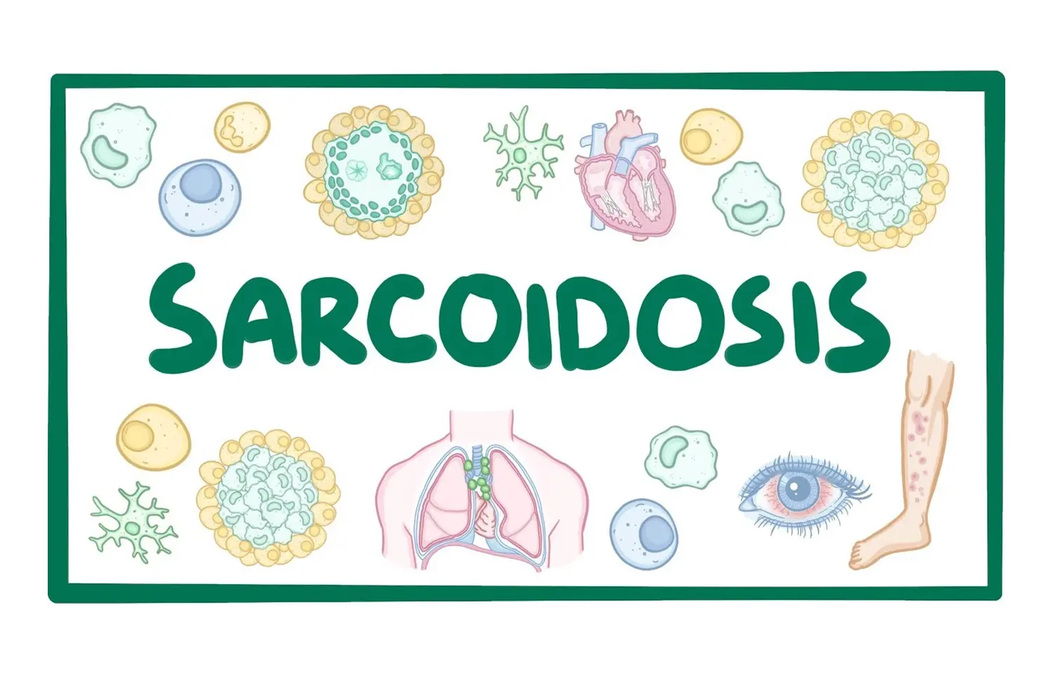
Sarcoidosis is a chronic inflammatory disease of unknown aetiology, characterised by the formation of non-caseating granulomata involving one or more organs. Extrathoracic manifestations of the disease can include otorhinolaryngological problems, with a particular predilection for upper and lower airway involvement. This article will outline the background of sarcoidosis, describe recognised ENT manifestations and discuss current treatment options.
Cereceda-Monteoliva performed a PubMed literature review to determine the evidence base supporting this.
Pathophysiology
The cause of sarcoidosis is not yet well understood, but an antigentriggered, cell-mediated immune response is known to initiate the sarcoidosis disease process. Genetic susceptibility and specific environmental or infectious triggers combine to perpetuate a chronic cell-mediated immunological response. T lymphocytes and macrophages accumulate and produce inflammatory mediators such as cytokines (particularly TNF-alpha, IL-12 and IL-18), which lead to the formation of granulomata. Certain HLA haplotypes are suggested to predispose to the disease and may be associated with disease phenotype and outcome, although evidence for this remains inconclusive.
Diagnosis
Sarcoidosis is diagnosed on the basis of clinical and radiological suspicion, combined with biopsy evidence of non-caseating granulomata, in the absence of any other cause of granulomatous disorders. Presenting symptoms vary widely and are non-specific; these include low-grade fever, unexpected weight loss, night sweats and arthralgias.
Imaging techniques routinely employed include chest radiography, where characteristic bilateral perihilar lymphadenopathy is often considered diagnostic, and high-resolution computed tomography (HRCT) in cases of atypical or complicated chest sarcoid. MRI (with gadolinium contrast) is commonly used for detection of cardiac involvement with sensitivity of 75%-100%. Nuclear imaging (with gallium) and 18F-fluorodeoxyglucose positron-emission tomography (FDG-PET) can also be used to evaluate inflammatory activity.
Biopsy typically yields multiple epithelioid cell granulomata made from mononuclear cells, with variable degrees of necrosis, leucocyte infiltration and hyaline fibrosis, with a reported diagnostic accuracy of 40-90% in transbronchial lung biopsy (TBLB), the gold standard for pulmonary sarcoidosis, now recommended above the transoesophageal and mediastinoscopic approaches. Supporting evidence for sarcoidosis can include a high CD4/CD8 T lymphocyte ratio on bronchoalveolar lavage, with a high specificity of 95% yet a low sensitivity of 52-59%. Blood tests can provide supporting evidence for the diagnosis of sarcoidosis. Serum angiotensin-converting enzyme (ACE) levels are raised in approximately two-thirds of patients with sarcoidosis; however, the non-specific and insensitive nature of this rise relegates its use to monitoring the course of disease. Hypercalcaemia may also be seen and other acute phase reactants, such as ESR, may also be raised.
General clinical manifestations
Intrathoracic manifestations of sarcoidosis include pulmonary infiltrates and hilar lymphadenopathy as well as cardiac sarcoidosis, which can be life-threatening. Extrathoracic disease occurs in around 50% of patients, and virtually every organ can be affected. The extent and degree of disease is extremely variable between patients. Skin, lymph node, eye and liver involvement are most common outside of the thorax. Around 5%-15% of patients with sarcoid have ENT manifestations of sarcoidosis which may be the presenting symptom of their disease. The differential often includes vasculitides such as granulomatosis with polyangiitis, formerly called Wegener's, or eosinophilic granulomatosis with polyangiitis, formerly Churg-Strauss syndrome, granulomas of infective origin (such as tuberculosis, aspergillosis or actinomycosis), and inflammatory diseases with extrasystemic manifestations, such as Crohn's disease, which should be excluded. Sinonasal sarcoidosis is rare, but has been described in numerous case studies, and is a well-recognised chronic and stubborn form of the disease. Sarcoidosis can also involve the larynx, salivary glands and ear in rare cases.
Treatment principles
Although existing guidelines are limited in scope, the BTS Clinical Statement on Pulmonary Sarcoidosis recommends medical management with oral corticosteroids in long-standing disease, with significantly increased daily dosages in more acute flare-ups.
Symptomatic relief and lifestyle modifications, including smoking cessation and psychological support, should also be offered as routine. The recommended second-line agent is methotrexate, orally or subcutaneously, and advanced pulmonary disease may warrant referral for lung transplantation. Anecdotally, steroids are also the mainstay of treatment in otolaryngologic disease. However, the risks of uncontrolled disease need to be balanced against the incumbent risks and side-effects of long-term corticosteroid therapy.
Adjunctive or alternate drug therapies have therefore also been used in order to reduce steroid doses. These include the following:
▪ Cytotoxic agents (such as methotrexate, azathioprine, cyclophosphamide and chlorambucil).
▪ Anti-malarial drugs (chloroquine and hydroxychloroquine).
▪ TNF-alpha inhibitors (infliximab, adalimumab, etanercept).
No control studies exist as yet comparing these therapies to steroid treatment, but reports of effective treatment exist in cases of sarcoidosis with extrathoracic manifestations such as lupus pernio and uveitis.
ENT manifestations are present in 5%-15% of patients with sarcoidosis, often as a presenting feature, and require vigilance for swift recognition and coordinated additional treatment specific to the organ.
Laryngeal sarcoidosis presents with difficulty in breathing, dysphonia and cough, and may be treated by speech and language therapy (SLT) or intralesional injection, dilatation or tissue reduction.
Nasal disease presents with crusting, rhinitis, nasal obstruction and anosmia, usually without sinus involvement. It is treated by topical nasal or intralesional treatments but may also require endoscopic sinus surgery, laser treatment or even nasal reconstruction.
Otological disease is uncommon but includes audiovestibular symptoms, both sensorineural and conductive hearing loss, and skin lesions.
Prognosis
Sarcoidosis generally shows spontaneous remission in 12-36 months (50%-60% of cases). Prognosis is usually favourable, although a 3%-9% mortality is reported, usually from lung or cardiac complications. It is difficult to recommend ENT-specific treatments given the scarcity of data that exists on the management and prognosis of site-specific manifestations.
The consequences of ENT manifestations of sarcoidosis can be uncomfortable, disabling and disfiguring and those affecting the larynx can be life-threatening. The coordinated management of these requires good diagnostic skills and multimodal treatment. Established treatments using corticosteroids are observed to be effective but not without risks of long-term use. Intralesional treatments are commonly reported, and surgical excision of tissue may be necessary. For clinical care to evolve and improve, it is vital for affected patients to be managed using all the resources available to the multidisciplinary team and that outcomes are studied and recorded so as to establish best practice in this area.
Key Points
• Sarcoidosis is a multisystemic inflammatory disease with ENT manifestations present in 5%-15% of patients, often as a presenting feature.
• Diagnosis typically requires evidence of non-caseating granulomata, and prognosis is favourable with treatment.
• "Turban-shaped" epiglottis and restricted glottic mobility are observed on endoscopic examination of laryngeal sarcoidosis, where treatments include speech and language therapy (SLT), intralesional injection, dilatation and tissue reduction surgery.
• Sinonasal sarcoidosis is staged according to location and severity of disease, which guides treatment options, including intranasal, intralesional and systemic treatments, separately or in combination with topical, nasal and intralesional corticosteroid and endoscopic sinus surgery.
• Otologic sarcoidosis is difficult to diagnose but responds well to oral or intralesional corticosteroid therapy.
Source: Cereceda-Monteoliva et al.; Clinical Otolaryngology. 2021;46:935–940.
DOI: 10.1111/coa.13814
Dr Ishan Kataria has done his MBBS from Medical College Bijapur and MS in Ophthalmology from Dr Vasant Rao Pawar Medical College, Nasik. Post completing MD, he pursuid Anterior Segment Fellowship from Sankara Eye Hospital and worked as a competent phaco and anterior segment consultant surgeon in a trust hospital in Bathinda for 2 years.He is currently pursuing Fellowship in Vitreo-Retina at Dr Sohan Singh Eye hospital Amritsar and is actively involved in various research activities under the guidance of the faculty.
Dr Kamal Kant Kohli-MBBS, DTCD- a chest specialist with more than 30 years of practice and a flair for writing clinical articles, Dr Kamal Kant Kohli joined Medical Dialogues as a Chief Editor of Medical News. Besides writing articles, as an editor, he proofreads and verifies all the medical content published on Medical Dialogues including those coming from journals, studies,medical conferences,guidelines etc. Email: drkohli@medicaldialogues.in. Contact no. 011-43720751


