- Home
- Medical news & Guidelines
- Anesthesiology
- Cardiology and CTVS
- Critical Care
- Dentistry
- Dermatology
- Diabetes and Endocrinology
- ENT
- Gastroenterology
- Medicine
- Nephrology
- Neurology
- Obstretics-Gynaecology
- Oncology
- Ophthalmology
- Orthopaedics
- Pediatrics-Neonatology
- Psychiatry
- Pulmonology
- Radiology
- Surgery
- Urology
- Laboratory Medicine
- Diet
- Nursing
- Paramedical
- Physiotherapy
- Health news
- Fact Check
- Bone Health Fact Check
- Brain Health Fact Check
- Cancer Related Fact Check
- Child Care Fact Check
- Dental and oral health fact check
- Diabetes and metabolic health fact check
- Diet and Nutrition Fact Check
- Eye and ENT Care Fact Check
- Fitness fact check
- Gut health fact check
- Heart health fact check
- Kidney health fact check
- Medical education fact check
- Men's health fact check
- Respiratory fact check
- Skin and hair care fact check
- Vaccine and Immunization fact check
- Women's health fact check
- AYUSH
- State News
- Andaman and Nicobar Islands
- Andhra Pradesh
- Arunachal Pradesh
- Assam
- Bihar
- Chandigarh
- Chattisgarh
- Dadra and Nagar Haveli
- Daman and Diu
- Delhi
- Goa
- Gujarat
- Haryana
- Himachal Pradesh
- Jammu & Kashmir
- Jharkhand
- Karnataka
- Kerala
- Ladakh
- Lakshadweep
- Madhya Pradesh
- Maharashtra
- Manipur
- Meghalaya
- Mizoram
- Nagaland
- Odisha
- Puducherry
- Punjab
- Rajasthan
- Sikkim
- Tamil Nadu
- Telangana
- Tripura
- Uttar Pradesh
- Uttrakhand
- West Bengal
- Medical Education
- Industry
Sarcoidosis of the ear, nose and throat: A review of the literature

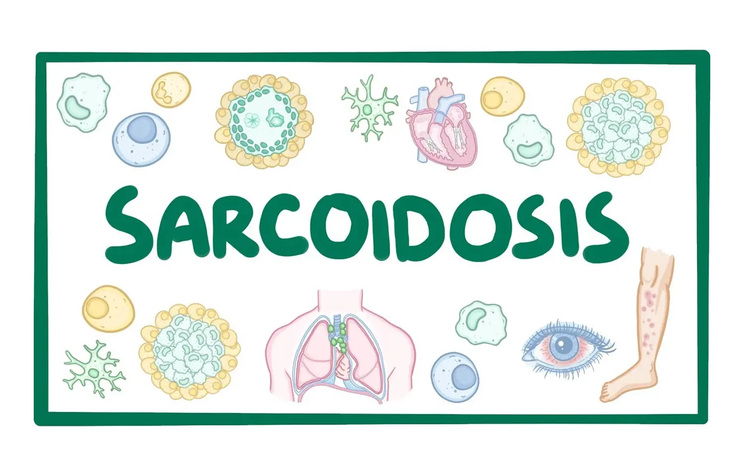
Sarcoidosis is a chronic inflammatory disease of unknown aetiology, characterised by the formation of non-caseating granulomata involving one or more organs. Extrathoracic manifestations of the disease can include otorhinolaryngological problems, with a particular predilection for upper and lower airway involvement. This article by CERECEDA-MONTEOLIVA et al outlines the background of sarcoidosis, describe recognised ENT manifestations and discuss current treatment options.
Authors performed a PubMed literature review to determine the evidence base supporting this.
Epidemiology
The prevalence of sarcoidosis varies geographically between 1 and 40 cases per 100 000 and is more prevalent in south-eastern United States and Scandinavia. It occurs more commonly in a bimodal distribution affecting the young and middle-aged populations, and females are affected more frequently than men and Black people more than White.
Pathophysiology
The cause of sarcoidosis is not yet well understood, but an antigen triggered, cell-mediated immune response is known to initiate the sarcoidosis disease process. Genetic susceptibility and specific environmental or infectious triggers combine to perpetuate a chronic cell-mediated immunological response. T lymphocytes and macrophages accumulate and produce inflammatory mediators such as cytokines (particularly TNF-alpha, IL-12 and IL-18), which lead to the formation of granulomata. Certain HLA haplotypes are suggested to predispose to the disease and may be associated with disease phenotype and outcome, although evidence for this remains inconclusive.
Diagnosis
Sarcoidosis is diagnosed on the basis of clinical and radiological suspicion, combined with biopsy evidence of non-caseating granulomata, in the absence of any other cause of granulomatous disorders, as stated in the Official American Thoracic Society (ATS) Clinical Practice Guideline for Diagnosis and Detection and the recent British Thoracic Society (BTS) Clinical Statement on Pulmonary Sarcoidosis.
Presenting symptoms vary widely and are non-specific; these include low-grade fever, unexpected weight loss, night sweats and arthralgias in up to 30% of patients. Traditionally, the Kveim reaction was an important clinical observation in making a diagnosis of sarcoidosis; however, its use has fallen out of favour.
Imaging techniques routinely employed include chest radiography, where characteristic bilateral perihilar lymphadenopathy is often considered diagnostic and high-resolution computed tomography (HRCT) in cases of atypical or complicated chest sarcoid. MRI (with gadolinium contrast) is commonly used for detection of cardiac involvement with sensitivity of 75%-100%. Nuclear imaging (with gallium) and 18F-fluorodeoxyglucose positron-emission tomography (FDG-PET) can also be used to evaluate inflammatory activity, the latter demonstrating a sensitivity of 88% and a specificity of only 39%.
Biopsy typically yields multiple epithelioid cell granulomata made from mononuclear cells, with variable degrees of necrosis, leucocyte infiltration and hyaline fibrosis, with a reported diagnostic accuracy of 40-90% in transbronchial lung biopsy (TBLB), the gold standard for pulmonary sarcoidosis, now recommended above the transoesophageal and mediastinoscopic approaches. Supporting evidence for sarcoidosis can include a high CD4/CD8 T lymphocyte ratio on bronchoalveolar lavage, with a high specificity of 95% yet a low sensitivity of 52-59%.
Blood tests can provide supporting evidence for the diagnosis of sarcoidosis. Serum angiotensin-converting enzyme (ACE) levels are raised in approximately two-thirds of patients with sarcoidosis; however, the non-specific and insensitive nature of this rise relegates its use to monitoring the course of disease. Hypercalcaemia may also be seen and other acute phase reactants, such as ESR, may also be raised.
General clinical manifestations
Intrathoracic manifestations of sarcoidosis include pulmonary infiltrates and hilar lymphadenopathy affecting 90% of patients as well as cardiac sarcoidosis (more common in older Japanese females), which can be life-threatening. Extrathoracic disease occurs in around 50% of patients, and virtually every organ can be affected. The extent and degree of disease is extremely variable between patients. Skin, lymph node, eye and liver involvement are most common outside of the thorax, each being found in between 10% and 25% of patients.
Around 5%-15% of patients with sarcoid have ENT manifestations of sarcoidosis which may be the presenting symptom of their disease. The differential often includes vasculitides such as granulomatosis with polyangiitis, formerly called Wegener's, or eosinophilic granulomatosis with polyangiitis, formerly Churg-Strauss syndrome, granulomas of infective origin (such as tuberculosis, aspergillosis or actinomycosis), and inflammatory diseases with extrasystemic manifestations, such as Crohn's disease, which should be excluded. Sinonasal sarcoidosis is rare (occurring in 1%-4% of patients), but has been described in numerous case studies, and is a well-recognised chronic and stubborn form of the disease. Sarcoidosis can also involve the larynx, salivary glands and ear in rare cases.
Treatment principles
Although existing guidelines are limited in scope, the BTS Clinical Statement on Pulmonary Sarcoidosis recommends medical management with oral corticosteroids in long-standing disease, with significantly increased daily dosages in more acute flare-ups. Symptomatic relief and lifestyle modifications, including smoking cessation and psychological support, should also be offered as routine. The recommended second-line agent is methotrexate, orally or subcutaneously, and advanced pulmonary disease may warrant referral for lung transplantation. Anecdotally, steroids are also the mainstay of treatment in otolaryngologic disease.
However, the risks of uncontrolled disease need to be balanced against the incumbent risks and side-effects of long-term corticosteroid therapy. Adjunctive or alternate drug therapies have therefore also been used in order to reduce steroid doses. These include the following:
▪ Cytotoxic agents (such as methotrexate, azathioprine, cyclophosphamide and chlorambucil).
▪ Anti-malarial drugs (chloroquine and hydroxychloroquine).
▪ TNF-alpha inhibitors (infliximab, adalimumab, etanercept).
- ENT MANIFESTATIONS OF DISEASE
Laryngeal sarcoidosis
Laryngeal sarcoidosis is particularly rare, with a large cohort of patients with systemic sarcoidosis demonstrating an incidence of only 0.6%. As with other manifestations of sarcoidosis, age at presentation is typically from 20 to 40 years old.
The presenting complaint in the majority of patients relates to difficulty breathing and less commonly dysphonia and cough. Pain is not usually a feature. Only a small proportion of patients complain of dysphagia and if so, it is rarely the sole symptom. Those with dysphagia inevitably modify their diet and some silently aspirate. Given the natural history and slow progression of this granulomatous disease, emergency department presentations are rare. Cases of emergency tracheostomy exist; however, most patients present with a gradual decline. Stridor is common but mild. Although symptom onset is gradual, the impact on quality of life is significant with a reduction in exercise capacity, compounding respiratory disease in affected young patients.
Endoscopic examination typically reveals supraglottic swelling and deformity, with the epiglottis, arytenoids and aryepiglottic folds involved. "Turban"-shaped epiglottis is typically described due to the resultant morphological change after inflammatory and granulomatous infiltrate. True vocal fold paresis is rare, but there can often be an appearance of restricted glottic mobility contributing to dysphonia due to the inflammatory process and resultant thickening.
Prior to a diagnosis of laryngeal sarcoidosis, a broad differential must be considered given that presenting symptoms related to airway and voice are also seen in many more common laryngeal pathologies including vocal fold paralysis, supraglottitis and laryngotracheal stenosis. The laryngeal appearance may resemble acute infection or other granulomatous diseases such as tuberculosis and vasculitis. Malignancy may rarely present in this way and must always be considered. It is crucial that the entire respiratory tract has been assessed, as more foci of disease suggest a systemic process such as sarcoidosis, where solitary lesions are more likely to be due to localised pathology. Investigations may be performed but only a biopsy demonstrating non-caseating granulomata is diagnostic.
Conservative options typically relate to speech and language therapy for patients with symptomatic dysphonia or dysphagia. Medical therapy for those with pre-existing sarcoidosis has often been initiated as described above. The majority of patients that require surgical intervention have problematic airway symptoms. Microlaryngoscopy and biopsy are performed for a definitive diagnosis, whilst also providing an opportunity to dilate the airway if required. There have been descriptions of intralesional steroid injection, as well as the use of mitomycin-C to prevent recurrent scar. In general, surgical techniques employ the CO2 laser for tissue reduction or excision—the addition of the previously described "pepper pot" technique has been demonstrated to reduce patients' Medical Research Council dyspnoea grade.
Sinonasal Sarcoidosis
Sinonasal involvement occurs in 1%-4% of sarcoidosis cases. Involvement tends to be mucosal but can involve the bone of the nose and paranasal sinuses. Typical symptoms include chronic crusting rhinitis (70%-90%), nasal obstruction (unilateral or bilateral) (80%-90%) and anosmia (70%). Epistaxis (2%) and nasal deformity can occur in advanced and destructive cases.
Clinical examination may reveal hypertrophic spots and a characteristic purplish colouring of the nasal mucosa, with granulations on the septum and inferior turbinates. Less commonly, the paranasal sinuses may be partially or completely opacified, with mucosal thickening and osteomeatal obstruction. There have also been cases of sinonasal sarcoidosis with peripheral nerve involvement, salivary gland extension and intracranial extension. The sinonasal cavity is commonly affected in vasculitic conditions and these are therefore the primary differential diagnosis, in addition to chronic rhinosinusitis, prior to a tissue diagnosis.
Treatment of sinonasal sarcoidosis depends on the location and severity of the disease. Hence, a staging system is described by Krepsi et al. Medical nasal, intralesional and systemic treatments have been tried separately and in combination with topical nasal and intralesional corticosteroid therapy having the benefit of avoiding the complications that occur with the use of systemic corticosteroids. However, if the symptoms and clinical destruction are severe, systemic corticosteroids, as outlined above, are indicated.
Surgical treatment can be effective where medical treatment has failed, and in particular in cases which develop anatomical blockage of sinus drainage pathways with sarcoidosis lesions. Endoscopic sinus surgery can markedly improve quality of life and reduce the need for systemic steroids, but it will not eradicate the disease or prevent recurrence. Successful laser surgery with a CO2 laser has been reported, as well as successful partial nasal reconstruction with a two-stage, right-sided paramedical forehead flap with a rib cartilage framework.
Staging of sinonasal sarcoidosis, as proposed by Krepsi et al
Stage 1 Mild, reversible disease without paranasal sinus involvement
Stage 2 Moderate, potentially reversible disease without sinus involvement
Stage 3 Moderate, potentially reversible disease with sinus involvement
Otologic sarcoidosis
Audiovestibular symptoms of sarcoidosis are uncommon but can include sensorineural hearing loss, vertigo, gait disturbance and disequilibrium from neurosarcoidosis affecting the vestibulocochlear nerves. Middle ear sarcoidosis has also been reported as a sentinel manifestation of the disease, presenting with tinnitus, hearing loss, purulent otorrhoea and otalgia. In this scenario, audiometric findings are mixed or conductive in nature, with CT demonstrating soft tissue masses in the tympanic cavity extending into the mastoid antrum and air cells. While skin is commonly involved in sarcoidosis, the external ear is seldom involved. Cases described include tender and non-tender raised nodules, swelling or inflammation that can affect the helix, lobules or external auditory canal.
Otologic symptoms usually improve with oral or intralesional corticosteroid therapy. Infliximab, a TNF-α blocking agent, has also been reported as successful in treating ear lobe sarcoidosis refractory to non-biologic immunosuppressants and intralesional injected corticosteroids. Case reports have suggested that surgical excision of granulation tissue alongside systemic steroid therapy can result in improvement of symptoms.
Prognosis
Sarcoidosis generally shows spontaneous remission in 12-36 months (50%-60% of cases). Prognosis is usually favourable, although a 3%-9% mortality is reported, usually from lung or cardiac complications. It is difficult to recommend ENT-specific treatments given the scarcity of data that exists on the management and prognosis of site-specific manifestations.
CONCLUSION
The consequences of ENT manifestations of sarcoidosis can be uncomfortable, disabling and disfiguring and those affecting the larynx can be life-threatening. The coordinated management of these requires good diagnostic skills and multimodal treatment. Established treatments using corticosteroids are observed to be effective but not without risks of long-term use. Intralesional treatments are commonly reported, and surgical excision of tissue may be necessary.
For clinical care to evolve and improve, it is vital for affected patients to be managed using all the resources available to the multidisciplinary team and that outcomes are studied and recorded so as to establish best practice in this area.
Source: CERECEDA-MONTEOLIVA et al; Clinical Otolaryngology. 2021;46:935–940.
DOI: 10.1111/coa.13814
Dr Ishan Kataria has done his MBBS from Medical College Bijapur and MS in Ophthalmology from Dr Vasant Rao Pawar Medical College, Nasik. Post completing MD, he pursuid Anterior Segment Fellowship from Sankara Eye Hospital and worked as a competent phaco and anterior segment consultant surgeon in a trust hospital in Bathinda for 2 years.He is currently pursuing Fellowship in Vitreo-Retina at Dr Sohan Singh Eye hospital Amritsar and is actively involved in various research activities under the guidance of the faculty.
Dr Kamal Kant Kohli-MBBS, DTCD- a chest specialist with more than 30 years of practice and a flair for writing clinical articles, Dr Kamal Kant Kohli joined Medical Dialogues as a Chief Editor of Medical News. Besides writing articles, as an editor, he proofreads and verifies all the medical content published on Medical Dialogues including those coming from journals, studies,medical conferences,guidelines etc. Email: drkohli@medicaldialogues.in. Contact no. 011-43720751


