- Home
- Medical news & Guidelines
- Anesthesiology
- Cardiology and CTVS
- Critical Care
- Dentistry
- Dermatology
- Diabetes and Endocrinology
- ENT
- Gastroenterology
- Medicine
- Nephrology
- Neurology
- Obstretics-Gynaecology
- Oncology
- Ophthalmology
- Orthopaedics
- Pediatrics-Neonatology
- Psychiatry
- Pulmonology
- Radiology
- Surgery
- Urology
- Laboratory Medicine
- Diet
- Nursing
- Paramedical
- Physiotherapy
- Health news
- Fact Check
- Bone Health Fact Check
- Brain Health Fact Check
- Cancer Related Fact Check
- Child Care Fact Check
- Dental and oral health fact check
- Diabetes and metabolic health fact check
- Diet and Nutrition Fact Check
- Eye and ENT Care Fact Check
- Fitness fact check
- Gut health fact check
- Heart health fact check
- Kidney health fact check
- Medical education fact check
- Men's health fact check
- Respiratory fact check
- Skin and hair care fact check
- Vaccine and Immunization fact check
- Women's health fact check
- AYUSH
- State News
- Andaman and Nicobar Islands
- Andhra Pradesh
- Arunachal Pradesh
- Assam
- Bihar
- Chandigarh
- Chattisgarh
- Dadra and Nagar Haveli
- Daman and Diu
- Delhi
- Goa
- Gujarat
- Haryana
- Himachal Pradesh
- Jammu & Kashmir
- Jharkhand
- Karnataka
- Kerala
- Ladakh
- Lakshadweep
- Madhya Pradesh
- Maharashtra
- Manipur
- Meghalaya
- Mizoram
- Nagaland
- Odisha
- Puducherry
- Punjab
- Rajasthan
- Sikkim
- Tamil Nadu
- Telangana
- Tripura
- Uttar Pradesh
- Uttrakhand
- West Bengal
- Medical Education
- Industry
Rare case of Pneumocephalus Due to a Bronchoatrial Fistula reported in NEJM

Dr Alan Zakko and Dr Hira Bakhtiar at Yale-New Haven Hospital, New Haven, CT have reported a rare case of Pneumocephalus due to a Bronchoatrial Fistula. The case has been published in the New England Journal of Medicine.
Pneumocephalus (PNC) is the presence of air in the intracranial cavity. The most frequent cause is trauma, but there are many other etiological factors, such as surgical procedures. PNC with compression of frontal lobes and the widening of the interhemispheric space between the tips of the frontal lobes is a characteristic radiological finding of the "Mount Fuji sign."
The incidence of pneumocephalus depends on the etiology and is seen in almost all post-craniotomy cases. The incidence following head injury varies depending on the series from 1% to as high as 82%.
A 61-year-old man with a history of stage IIIB squamous-cell carcinoma of the lung began to have seizurelike activity while he was undergoing restaging computed tomography (CT) in the hospital. He had a known primary cavitary lesion in the right lower lobe and had previously received three cycles of carboplatin and nanoparticle albumin-bound paclitaxel (Abraxane).
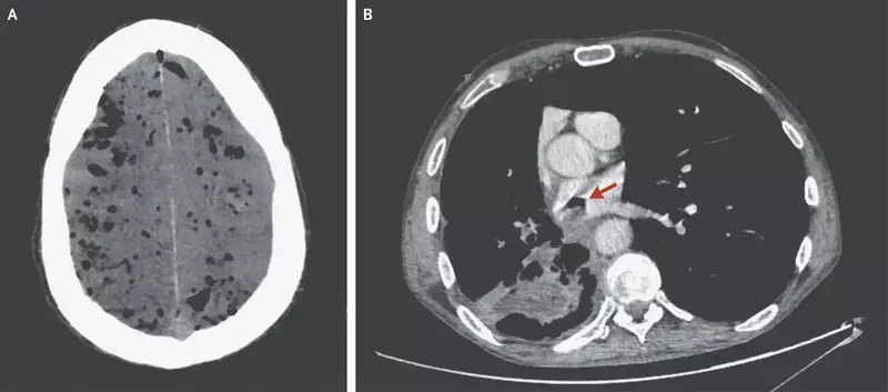
He was transported to the emergency department. Sudden respiratory failure and confusion developed, which resulted in emergency intubation. CT of the head revealed diffuse pneumocephalus (Panel A). The restaging CT scan of the chest showed that the previously identified cavitary lesion had formed a fistula between a bronchus in the right lower lobe and the left atrium (Panel B, arrow).
The pneumocephalus was thought to have been due to an air embolism that had originated from the bronchoatrial fistula. The patient later died in the intensive care unit.
N Engl J Med 2020; 382:1942
Dr Kamal Kant Kohli-MBBS, DTCD- a chest specialist with more than 30 years of practice and a flair for writing clinical articles, Dr Kamal Kant Kohli joined Medical Dialogues as a Chief Editor of Medical News. Besides writing articles, as an editor, he proofreads and verifies all the medical content published on Medical Dialogues including those coming from journals, studies,medical conferences,guidelines etc. Email: drkohli@medicaldialogues.in. Contact no. 011-43720751


