- Home
- Medical news & Guidelines
- Anesthesiology
- Cardiology and CTVS
- Critical Care
- Dentistry
- Dermatology
- Diabetes and Endocrinology
- ENT
- Gastroenterology
- Medicine
- Nephrology
- Neurology
- Obstretics-Gynaecology
- Oncology
- Ophthalmology
- Orthopaedics
- Pediatrics-Neonatology
- Psychiatry
- Pulmonology
- Radiology
- Surgery
- Urology
- Laboratory Medicine
- Diet
- Nursing
- Paramedical
- Physiotherapy
- Health news
- Fact Check
- Bone Health Fact Check
- Brain Health Fact Check
- Cancer Related Fact Check
- Child Care Fact Check
- Dental and oral health fact check
- Diabetes and metabolic health fact check
- Diet and Nutrition Fact Check
- Eye and ENT Care Fact Check
- Fitness fact check
- Gut health fact check
- Heart health fact check
- Kidney health fact check
- Medical education fact check
- Men's health fact check
- Respiratory fact check
- Skin and hair care fact check
- Vaccine and Immunization fact check
- Women's health fact check
- AYUSH
- State News
- Andaman and Nicobar Islands
- Andhra Pradesh
- Arunachal Pradesh
- Assam
- Bihar
- Chandigarh
- Chattisgarh
- Dadra and Nagar Haveli
- Daman and Diu
- Delhi
- Goa
- Gujarat
- Haryana
- Himachal Pradesh
- Jammu & Kashmir
- Jharkhand
- Karnataka
- Kerala
- Ladakh
- Lakshadweep
- Madhya Pradesh
- Maharashtra
- Manipur
- Meghalaya
- Mizoram
- Nagaland
- Odisha
- Puducherry
- Punjab
- Rajasthan
- Sikkim
- Tamil Nadu
- Telangana
- Tripura
- Uttar Pradesh
- Uttrakhand
- West Bengal
- Medical Education
- Industry
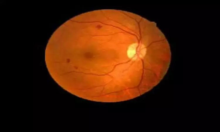
"Alone laser vs bevacizumab plus laser for diffuse diabetic macular edema" - ALBA randomized trial

Anti-vascular endothelial growth factor (antiVEGF) therapies have been tested as an alternative treatment to laser for diabetic macular edema (DME), showing such good results that they have become the first choice treatment.
Bevacizumab is a humanized full-length monoclonal antibody which encompasses all VEGF isoforms and has been used as anti-VEGF therapy for DME. Although not currently approved by the US Food and Drug Administration (FDA) for intraocular use, the injection of bevacizumab into the vitreous cavity has been performed without significant intraocular toxicity.
Alicia Pareja-Ríos et al carried out a study to determine the efficacy and safety of the off label intravitreal antiVEFG bevacizumab combined with grid laser photocoagulation compared with laser treatment alone in patients with diffuse DME over the course of 12months published in Therapeutic Advances in Ophthalmology.
"One of the main issues of intravitreal anti-VEGF therapy is the need for repeated injections. So, it has been combined with laser looking for an additive synergistic effect and the possibility of reducing the number of intravitreal injections. It had been thought that first thinning the retina with pharmacotherapy might enable better penetration of laser light to the retinal pigment epithelium and could improve the efficacy of laser. This strategy could reduce not only the number of injections but also the atrophy and the secondary effects induced by laser coagulation."
In this single-center randomized prospective, 12-month, phase III, randomized, independent controlled trial conducted in patients with diffuse DME, 32 patients were randomized to G1 (n = 15) or G2 (n = 17). In G1, laser was given at baseline and then pro re nata (PRN). In G2, three intravitreal bevacizumab (1.25mg) injections were given once every 6 weeks, then laser and then PRN. Analysis was performed by treatment as administered.
One eye of each patient was selected and treated as the study eye. If both eyes were eligible, the eye with the worse best corrected visual acuity (BCVA) assessed at the first visit was selected as the study eye.
Study participants were assigned randomly, using the opaque envelope method, to 1 of the 2 treatment arms.
The primary outcome measurement was the BCVA, while secondary outcomes included central foveal thickness (CFT) and macular volume.
Follow-up was planned for 1year. During that year, follow-up visits occurred every 4weeks (±1week) in G2 group, while in G1 group, visits were fixed by standard protocol after the retinal laser treatment during a period of 6months and every 4weeks (±1week) after 6months.
Safety evaluations, measurement of BCVA, eye examinations, and SD-OCT scans were performed at all follow-up visits.
Fluorescein angiography was performed at baseline at 6, and 12 months.
Measurements of glycosylated hemoglobin were obtained at baseline and at 3, 6, and 12months. Hematology and blood chemistry tests were performed at baseline and at 6 and 12months.
Finally, health-related quality of life, assessed through the National Eye Institute Visual Function Questionnaire (NEI VFQ-25) was applied at 12months in both groups.
Visual acuity
After treatments, BCVA in G2 was statistically higher (p value⩽0.01) than G1 in all follow-up visits.
Regarding statistical significance, the proportion of patients with visual improvement was statistically superior in G2 compared to G1 at 3 and 6 months, while treatment with laser therapy does not reach this value throughout the follow-up.
Central foveal thickness
The mean CFT was similar between groups at the beginning of the study (p=0.99). Both groups did not show any significant difference in CFT at month 3, 9, and 12. However, at month 6, G2 showed statistically lower values than G1.
Macular volume
There was no significant difference in average macular volume between groups during the study. Any of the groups achieved a normal macular volume during the study.
However, when the change in macular volume with respect to baseline was specifically analyzed, a significant reduction of macular volume in G2 was observed over time (p <0.05), whereas this fact did not occur in G1.
Type of DME
According to the classification of Panozzo and colleagues, DME was divided into three sub-groups: spongiform (E1), cyst (E2), and subretinal fluid (E3). In G1 (n = 15), there were 3 (20%), 7 (47%), and 5 (33%) patients with E1, E2, and E3, respectively. In G2 (n = 17), there were 0 (0%), 9 (53%), and 8 (47%), respectively.
There were significative differences between groups in patients with DME type E2, where G2 showed better values. Analyzing intra-group differences (e.g. change with respect baseline for a certain group), researchers found significant improvements only for G2 with DME type E2 at all follow-up visits.-
Regarding side-effects, the increasing use of bevacizumab is raising concerns about safety with long-term use of these agents. This drug has the potential to inhibit all the isoforms of VEGF-A and, in consequence, all its functions such as wound healing. The study did not find significant differences between both treatments regarding systemic safety.
The main limitation of this study is that it does not compare the response to bevacizumab alone, along with the two other arms. So it is difficult to assess whether the improvement in BCVA is due to the combined laser and bevacizumab or the latter alone. Additionally, the G2 group inferences may be the result of confounding bias created by the fact that a standalone bevacizumab group was not used to establish the efficacy of bevacizumab combined with focal laser used as adjunct following the loading dose.
Main strength of the study was it included only diffuse DME and a high percentage of naive patients suggesting that this type of patient may require a smaller number of intravitreal treatments.
In conclusion,the study suggests, "Laser combined with intravitreal bevacizumab was an effective option in the management of diffuse DME with central involvement through the follow-up. Results suggested that after 6months of follow-up, the combined treatment was superior to the laser, and it was maintained during the rest of the follow-up, even with the low number of injections reported in this study."
Source: Alicia Pareja-Ríos , Elena de Armas-Ramos, Ana Aldea-Perona and Sergio Bonaque-González Ther Adv Ophthalmol 2021, Vol. 13: 1–14
DOI: 10.1177/ 2515841420988210
Dr Ishan Kataria has done his MBBS from Medical College Bijapur and MS in Ophthalmology from Dr Vasant Rao Pawar Medical College, Nasik. Post completing MD, he pursuid Anterior Segment Fellowship from Sankara Eye Hospital and worked as a competent phaco and anterior segment consultant surgeon in a trust hospital in Bathinda for 2 years.He is currently pursuing Fellowship in Vitreo-Retina at Dr Sohan Singh Eye hospital Amritsar and is actively involved in various research activities under the guidance of the faculty.
Dr Kamal Kant Kohli-MBBS, DTCD- a chest specialist with more than 30 years of practice and a flair for writing clinical articles, Dr Kamal Kant Kohli joined Medical Dialogues as a Chief Editor of Medical News. Besides writing articles, as an editor, he proofreads and verifies all the medical content published on Medical Dialogues including those coming from journals, studies,medical conferences,guidelines etc. Email: drkohli@medicaldialogues.in. Contact no. 011-43720751


