- Home
- Medical news & Guidelines
- Anesthesiology
- Cardiology and CTVS
- Critical Care
- Dentistry
- Dermatology
- Diabetes and Endocrinology
- ENT
- Gastroenterology
- Medicine
- Nephrology
- Neurology
- Obstretics-Gynaecology
- Oncology
- Ophthalmology
- Orthopaedics
- Pediatrics-Neonatology
- Psychiatry
- Pulmonology
- Radiology
- Surgery
- Urology
- Laboratory Medicine
- Diet
- Nursing
- Paramedical
- Physiotherapy
- Health news
- Fact Check
- Bone Health Fact Check
- Brain Health Fact Check
- Cancer Related Fact Check
- Child Care Fact Check
- Dental and oral health fact check
- Diabetes and metabolic health fact check
- Diet and Nutrition Fact Check
- Eye and ENT Care Fact Check
- Fitness fact check
- Gut health fact check
- Heart health fact check
- Kidney health fact check
- Medical education fact check
- Men's health fact check
- Respiratory fact check
- Skin and hair care fact check
- Vaccine and Immunization fact check
- Women's health fact check
- AYUSH
- State News
- Andaman and Nicobar Islands
- Andhra Pradesh
- Arunachal Pradesh
- Assam
- Bihar
- Chandigarh
- Chattisgarh
- Dadra and Nagar Haveli
- Daman and Diu
- Delhi
- Goa
- Gujarat
- Haryana
- Himachal Pradesh
- Jammu & Kashmir
- Jharkhand
- Karnataka
- Kerala
- Ladakh
- Lakshadweep
- Madhya Pradesh
- Maharashtra
- Manipur
- Meghalaya
- Mizoram
- Nagaland
- Odisha
- Puducherry
- Punjab
- Rajasthan
- Sikkim
- Tamil Nadu
- Telangana
- Tripura
- Uttar Pradesh
- Uttrakhand
- West Bengal
- Medical Education
- Industry
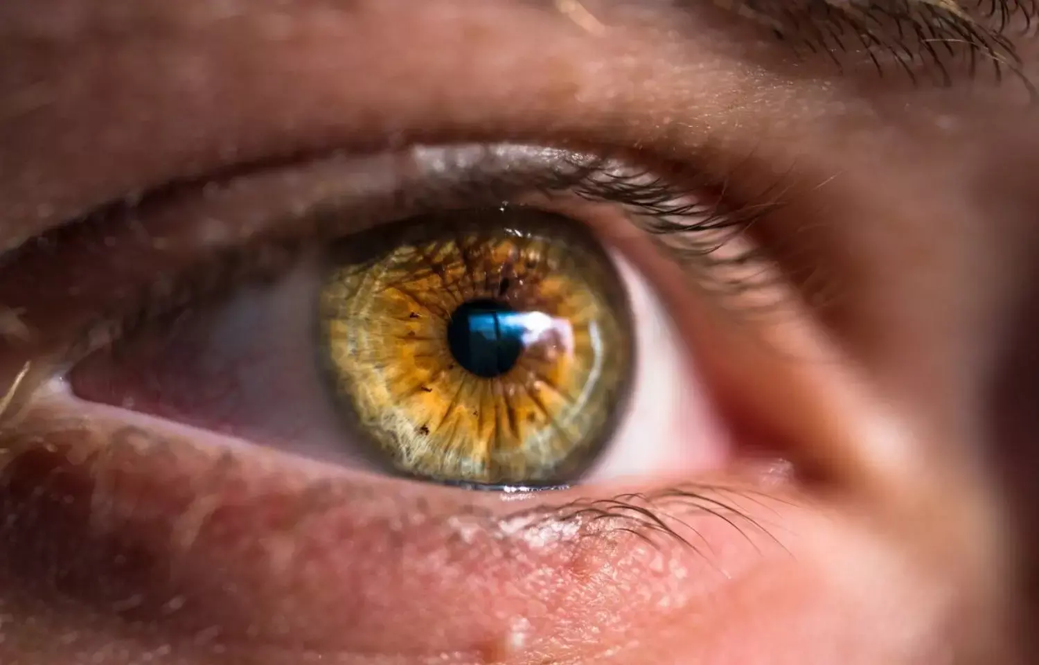
Green filter overlays make it easier to spot retinal cracks

New research found that the application of green filter overlay helps resident physicians to identify breaks on fundus photos when compared to traditional settings. The study results were published in the journal Graefe's Archive for Clinical and Experimental Ophthalmology.
Ophthalmologists can now more clearly see and record retinal diseases due to advancements in retinal imaging. The first ultra-widefield (UWF) fundus camera, the Optos confocal scanning laser ophthalmoscopy (cSLO; Optos PLC, Dunfermline, Scotland), allows for 200-degree fundus imaging in a single frame. Advances created in optos technology like pseudo color images produced by combining concurrently scanning red (633 nm) and green (532 nm) laser sources have revolutionized imaging techniques. Previous literature has shown that application of a green filter laser may be helpful in the assessment of the integrity of the retina. Hence, researchers conducted a study to assess the ability of resident physicians to identify retinal breaks on ultra-widefield color fundus photos using the traditional image compared to an image with a green filter overlay.
A retinal tear or hole observed in 10 eyes in the fundus photographs was shown to resident physicians. Each image was displayed to participants twice: once in its traditional color settings and once with a green filter applied. Participants were timed and given points for accurately identifying the break and also the time duration that they took to identify the pathology.
Findings:
- Residents were able to correctly identify more retinal breaks on fundus photos with a green filter overlay compared to photos with traditional settings (P = 0.02).
- Residents were also able to identify breaks on fundus photos more quickly on images with a green filter overlay compared to the traditional images (P < 0.001).
Thus, the green filter overlay has the potential for clinical use and helps resident physicians easily identify retinal breaks. It can also be used especially in telemedicine, to screen, diagnose, and monitor major eye diseases for patients in primary care and community settings.
Further reading: Moon, J.Y., Wai, K.M., Patel, N.S. et al. Visualization of retinal breaks on ultra-widefield fundus imaging using a digital green filter. Graefes Arch Clin Exp Ophthalmol 261, 935–940 (2023). https://doi.org/10.1007/s00417-022-05855-8
BDS, MDS
Dr.Niharika Harsha B (BDS,MDS) completed her BDS from Govt Dental College, Hyderabad and MDS from Dr.NTR University of health sciences(Now Kaloji Rao University). She has 4 years of private dental practice and worked for 2 years as Consultant Oral Radiologist at a Dental Imaging Centre in Hyderabad. She worked as Research Assistant and scientific writer in the development of Oral Anti cancer screening device with her seniors. She has a deep intriguing wish in writing highly engaging, captivating and informative medical content for a wider audience. She can be contacted at editorial@medicaldialogues.in.
Dr Kamal Kant Kohli-MBBS, DTCD- a chest specialist with more than 30 years of practice and a flair for writing clinical articles, Dr Kamal Kant Kohli joined Medical Dialogues as a Chief Editor of Medical News. Besides writing articles, as an editor, he proofreads and verifies all the medical content published on Medical Dialogues including those coming from journals, studies,medical conferences,guidelines etc. Email: drkohli@medicaldialogues.in. Contact no. 011-43720751


