- Home
- Medical news & Guidelines
- Anesthesiology
- Cardiology and CTVS
- Critical Care
- Dentistry
- Dermatology
- Diabetes and Endocrinology
- ENT
- Gastroenterology
- Medicine
- Nephrology
- Neurology
- Obstretics-Gynaecology
- Oncology
- Ophthalmology
- Orthopaedics
- Pediatrics-Neonatology
- Psychiatry
- Pulmonology
- Radiology
- Surgery
- Urology
- Laboratory Medicine
- Diet
- Nursing
- Paramedical
- Physiotherapy
- Health news
- Fact Check
- Bone Health Fact Check
- Brain Health Fact Check
- Cancer Related Fact Check
- Child Care Fact Check
- Dental and oral health fact check
- Diabetes and metabolic health fact check
- Diet and Nutrition Fact Check
- Eye and ENT Care Fact Check
- Fitness fact check
- Gut health fact check
- Heart health fact check
- Kidney health fact check
- Medical education fact check
- Men's health fact check
- Respiratory fact check
- Skin and hair care fact check
- Vaccine and Immunization fact check
- Women's health fact check
- AYUSH
- State News
- Andaman and Nicobar Islands
- Andhra Pradesh
- Arunachal Pradesh
- Assam
- Bihar
- Chandigarh
- Chattisgarh
- Dadra and Nagar Haveli
- Daman and Diu
- Delhi
- Goa
- Gujarat
- Haryana
- Himachal Pradesh
- Jammu & Kashmir
- Jharkhand
- Karnataka
- Kerala
- Ladakh
- Lakshadweep
- Madhya Pradesh
- Maharashtra
- Manipur
- Meghalaya
- Mizoram
- Nagaland
- Odisha
- Puducherry
- Punjab
- Rajasthan
- Sikkim
- Tamil Nadu
- Telangana
- Tripura
- Uttar Pradesh
- Uttrakhand
- West Bengal
- Medical Education
- Industry
Increased choroidal thickness in macular commotio retinae tied to decreased visual acuity

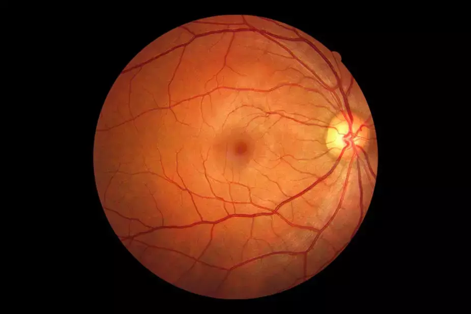
Commotio retinae, also known as Berlin edema, is a retina finding characterized by a transient, discrete sheen-like whitening of the retina seen after blunt ocular trauma. Optical coherence tomography (OCT) analysis of commotio retinae has shown characteristic morphologic changes, including hyporeflectivity of the cone outer segment tips and inner/outer segment junction as well as disruption of the external limiting membrane in cases with poorer visual prognosis. With advances in retinal imaging modalities such as spectral domain (SD) OCT with enhanced-depth imaging (EDI) protocols, the choroidal anatomy in many ophthalmic conditions is becoming better characterized.
Marie Burke et al carried out a study to investigate the subfoveal choroidal thickness (CT) and choroidal area (CA) in patients with unilateral commotio retinae of the macula. After analysis of SD-OCT images obtained with EDI protocol, CT, CA, and best-corrected visual acuity (BCVA) were compared using the nontraumatized fellow eye as the control.
This was a retrospective review of 16 eyes of 8 consecutive patients with unilateral macular commotio retinae within 7 days of blunt ocular trauma that underwent optical coherence tomography with enhanced-depth imaging seen at the institution. The contralateral, nontraumatized eye served as the control group.
All patients underwent spectral domain optical coherence tomography imaging with enhanced-depth imaging protocol. Using the electronic caliper within the Zeiss optical coherence tomography review software, CT was measured from the outer portion of the retinal pigment epithelium band to the inner surface of the sclera. The central horizontal and vertical rasters were averaged to calculate the final CT measurement of each eye. The final CA reading of each eye was obtained by averaging the central 1,500 mm2 of subfoveal CA using the same rasters. The researchers compared the CT, CA, and best-corrected visual acuity in traumatized eyes with macular commotio with their fellow nontraumatized control eyes.
RESULTS:
- The BCVA in traumatized eyes ranged from 20/30 to no light perception compared with a BCVA of 20/20 to 20/60 in control eyes.
- The difference in mean visual acuity was significant between traumatized eyes compared with control eyes (P = 0.0180).
- Eyes with macular commotio retinae displayed increased subfoveal CT (P = 0.0027).
- In addition, subfoveal mean CA was larger in eyes with macular commotio retinae compared with control eyes (P = 0.0279)
Several mechanisms for the sheen-like, opacified appearance of the retina in commotio retinae after blunt ocular trauma have been proposed, including extracellular edema, glial swelling, and photoreceptor outer segment disruption. Optical coherence tomography has dramatically improved understanding of the morphologic changes to the outer retina when commotio retinae involves the macula. In addition, macular photoreceptor function has been shown to be impaired in eyes with Berlin edema when tested with multifocal electroretinography.
Subfoveal CT and area were greater in eyes with commotio retinae than in the fellow nontraumatized eye after recent blunt trauma. Therefore, the commotio retinae does not only result in well-established changes to the outer retina but choroidal changes as well. The mechanism of increased CT is unclear; however the authors suggest that the findings could be attributable to choroidal vascular dilation in response to trauma.
Given the lack of automated CT and area measurements on existing OCT analysis software, one limitation of this study was the dependence on manual measurements of the CT using the software's built-in caliper function. As a result, intragrader variability may occur regarding recognition of choroidal boundaries. In an attempt to minimize the impact of this variability, CA was also analyzed in addition to CT.
The researchers concluded, "Subfoveal CT and CA were greater in eyes with commotio retinae when compared with normal fellow eyes. Increased CT and CA in macular commotio retinae were associated with decreased visual acuity."
Source: Marie Burke, Philip Lieu, Gary Abrams & Joseph Boss; RETINAL CASES & BRIEF REPORTS 15:417–420, 2021
Dr Ishan Kataria has done his MBBS from Medical College Bijapur and MS in Ophthalmology from Dr Vasant Rao Pawar Medical College, Nasik. Post completing MD, he pursuid Anterior Segment Fellowship from Sankara Eye Hospital and worked as a competent phaco and anterior segment consultant surgeon in a trust hospital in Bathinda for 2 years.He is currently pursuing Fellowship in Vitreo-Retina at Dr Sohan Singh Eye hospital Amritsar and is actively involved in various research activities under the guidance of the faculty.
Dr Kamal Kant Kohli-MBBS, DTCD- a chest specialist with more than 30 years of practice and a flair for writing clinical articles, Dr Kamal Kant Kohli joined Medical Dialogues as a Chief Editor of Medical News. Besides writing articles, as an editor, he proofreads and verifies all the medical content published on Medical Dialogues including those coming from journals, studies,medical conferences,guidelines etc. Email: drkohli@medicaldialogues.in. Contact no. 011-43720751


