- Home
- Medical news & Guidelines
- Anesthesiology
- Cardiology and CTVS
- Critical Care
- Dentistry
- Dermatology
- Diabetes and Endocrinology
- ENT
- Gastroenterology
- Medicine
- Nephrology
- Neurology
- Obstretics-Gynaecology
- Oncology
- Ophthalmology
- Orthopaedics
- Pediatrics-Neonatology
- Psychiatry
- Pulmonology
- Radiology
- Surgery
- Urology
- Laboratory Medicine
- Diet
- Nursing
- Paramedical
- Physiotherapy
- Health news
- Fact Check
- Bone Health Fact Check
- Brain Health Fact Check
- Cancer Related Fact Check
- Child Care Fact Check
- Dental and oral health fact check
- Diabetes and metabolic health fact check
- Diet and Nutrition Fact Check
- Eye and ENT Care Fact Check
- Fitness fact check
- Gut health fact check
- Heart health fact check
- Kidney health fact check
- Medical education fact check
- Men's health fact check
- Respiratory fact check
- Skin and hair care fact check
- Vaccine and Immunization fact check
- Women's health fact check
- AYUSH
- State News
- Andaman and Nicobar Islands
- Andhra Pradesh
- Arunachal Pradesh
- Assam
- Bihar
- Chandigarh
- Chattisgarh
- Dadra and Nagar Haveli
- Daman and Diu
- Delhi
- Goa
- Gujarat
- Haryana
- Himachal Pradesh
- Jammu & Kashmir
- Jharkhand
- Karnataka
- Kerala
- Ladakh
- Lakshadweep
- Madhya Pradesh
- Maharashtra
- Manipur
- Meghalaya
- Mizoram
- Nagaland
- Odisha
- Puducherry
- Punjab
- Rajasthan
- Sikkim
- Tamil Nadu
- Telangana
- Tripura
- Uttar Pradesh
- Uttrakhand
- West Bengal
- Medical Education
- Industry
Multifocal Epitheloid Hemangioma of Bone-A Rare Entity

Epithelioid hemangiomas (EHs) are rare vascular lesions which generally affect the skin and subcutaneous tissue but rarely seen in bones. It is a benign entity but intermediate grade, i.e., locally aggressive in nature. It has very confusing clinicoradiological and histopathological features which make diagnosis difficult and help us to avoid inappropriate treatment.
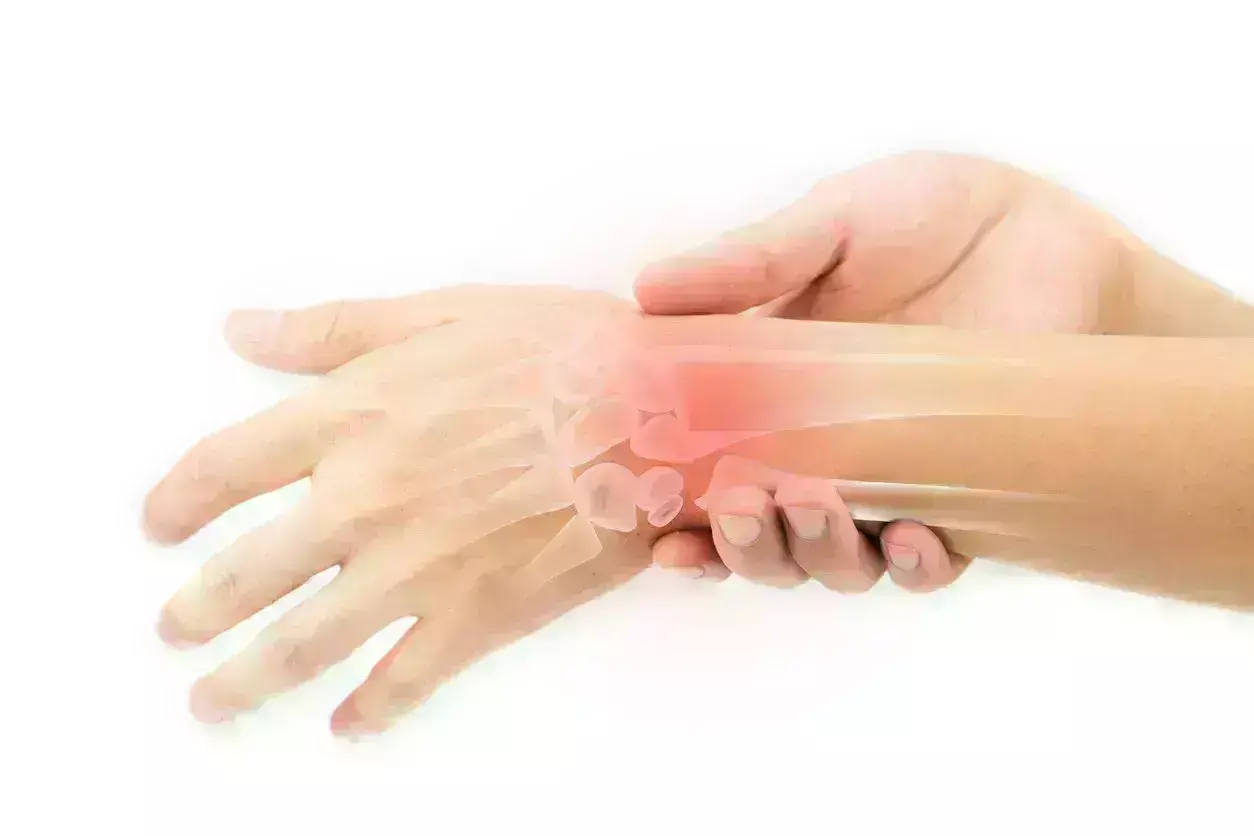
A 36-year-old male presented with pain and swelling over the right wrist extending toward the dorsal aspect of the hand associated with difficulty in wrist range of movements, for the past 3 months. There was no history of trauma or any twisting injury and no history of any fever. The swelling did not respond to any analgesics. Moreover, the swelling was increasing day by day, but there were no erythematous changes over the skin. Upon examination, there was tenderness over the wrist joint and carpometacarpal joints with a restricted range of movements of the wrist and multiple lobulated swelling felt over the dorsal aspect of the wrist.
After initial examination, the patient reported with X-ray and magnetic resonance imaging (MRI). A plain X-ray of the wrist showed a destructive lytic lesion over the distal radius which had ill-defined margins, lytic lesions also seen in the base of the 2nd and 3rd metacarpal base. MRI report showed an osteolytic lesion measuring 2.5 × 2.4 cm distal end of radius extending to a subarticular location with extraosseous soft-tissue component breaching the volar surface of distal radius. Multiple lytic lesions involving the trapezium, trapezoid bone, and capitate bones with associated marrow edema lytic lesions are also seen in the base of 2nd and 3rd metacarpals with enhancing soft tissue lesion measuring 2.9 × 2.1 cm abutting the carpal bones also seen. This was followed by whole-body positron emission tomography-computed tomography (CT) scan which revealed increased fluorodeoxyglucose uptake of standardized uptake value Max-5 in the distal radius with soft-tissue component and involvement of multiple carpal bones as described in MRI reports.
The authors performed a closed Jamshedji needle biopsy which was reported as EH. Then, he was managed with extended curettage + bone grafting + bone cementing and plating.
The patient was kept in close follow-up and there was no recurrence till 1-year post-operative period.
The authors concluded that – “EH of bone is a rare tumor and has a difficult diagnosis due to its aggressive clinicoradiological features. This case had aggressive radiological features, extensive soft tissue components, and multifocality which were looking like a malignant bone tumor with extensive soft-tissue involvement. Only after a closed J needle biopsy, expert histopathological study, and clinicoradiological correlation helped us to come to a confirmed diagnosis of EH. The case highlights clinical, radiological, and histopathological findings of EH of bone and also provides insights about the approach to these uncommon locally aggressive bone tumors. This case will help orthopedic surgeons and orthopedic onco-surgeons about the diagnostic approach and management of these rare bone tumors. An expert histopathological examination with the judicious help of an IHC panel is essential for a proper diagnosis which helps onco-pathologists and pathologists to understand the nature of the disease. Finally, coordination between orthopedic surgeons, radiologists, and pathologists (O.R.P) teams is a key for diagnosing rare bone tumors and its management.”
Further reading:
Multifocal Epitheloid Hemangioma of the Bone – A Rare Entity Bibhudutta Malla et al Journal of Orthopaedic Case Reports 2024 November:14(11):4-9 https://doi.org/10.13107/jocr.2024.v14.i11.4894
MBBS, Dip. Ortho, DNB ortho, MNAMS
Dr Supreeth D R (MBBS, Dip. Ortho, DNB ortho, MNAMS) is a practicing orthopedician with interest in medical research and publishing articles. He completed MBBS from mysore medical college, dip ortho from Trivandrum medical college and sec. DNB from Manipal Hospital, Bengaluru. He has expirence of 7years in the field of orthopedics. He has presented scientific papers & posters in various state, national and international conferences. His interest in writing articles lead the way to join medical dialogues. He can be contacted at editorial@medicaldialogues.in.


