- Home
- Medical news & Guidelines
- Anesthesiology
- Cardiology and CTVS
- Critical Care
- Dentistry
- Dermatology
- Diabetes and Endocrinology
- ENT
- Gastroenterology
- Medicine
- Nephrology
- Neurology
- Obstretics-Gynaecology
- Oncology
- Ophthalmology
- Orthopaedics
- Pediatrics-Neonatology
- Psychiatry
- Pulmonology
- Radiology
- Surgery
- Urology
- Laboratory Medicine
- Diet
- Nursing
- Paramedical
- Physiotherapy
- Health news
- Fact Check
- Bone Health Fact Check
- Brain Health Fact Check
- Cancer Related Fact Check
- Child Care Fact Check
- Dental and oral health fact check
- Diabetes and metabolic health fact check
- Diet and Nutrition Fact Check
- Eye and ENT Care Fact Check
- Fitness fact check
- Gut health fact check
- Heart health fact check
- Kidney health fact check
- Medical education fact check
- Men's health fact check
- Respiratory fact check
- Skin and hair care fact check
- Vaccine and Immunization fact check
- Women's health fact check
- AYUSH
- State News
- Andaman and Nicobar Islands
- Andhra Pradesh
- Arunachal Pradesh
- Assam
- Bihar
- Chandigarh
- Chattisgarh
- Dadra and Nagar Haveli
- Daman and Diu
- Delhi
- Goa
- Gujarat
- Haryana
- Himachal Pradesh
- Jammu & Kashmir
- Jharkhand
- Karnataka
- Kerala
- Ladakh
- Lakshadweep
- Madhya Pradesh
- Maharashtra
- Manipur
- Meghalaya
- Mizoram
- Nagaland
- Odisha
- Puducherry
- Punjab
- Rajasthan
- Sikkim
- Tamil Nadu
- Telangana
- Tripura
- Uttar Pradesh
- Uttrakhand
- West Bengal
- Medical Education
- Industry
AI Model Accurately Detects Low Bone Density from Chest X-Rays: Study

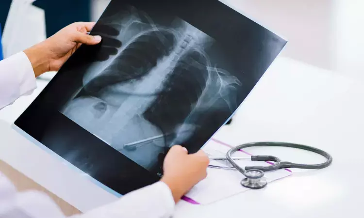
A new study published in Academic Radiology has demonstrated that an artificial intelligence (AI)-based model can accurately detect low bone mineral density (BMD) from routine chest radiographs, potentially offering a noninvasive tool for early screening and intervention.
The AI model was trained on thousands of chest X-rays and corresponding dual-energy X-ray absorptiometry (DXA) scans, the current gold standard for measuring BMD. Researchers found that the AI system achieved high sensitivity and specificity in identifying patients at risk for osteoporosis, particularly in detecting bone loss in the lumbar vertebrae.
In addition to classification, the model was capable of highlighting regions of low density directly on the radiographs, improving interpretability for clinicians. These findings suggest that integrating AI algorithms into routine imaging workflows could help identify at-risk individuals during unrelated chest imaging, thus enabling earlier diagnosis and targeted preventive strategies before significant bone loss or fractures occur.
The study emphasizes that because chest X-rays are one of the most commonly performed imaging tests globally, embedding such AI-driven tools could have a far-reaching impact on osteoporosis detection, especially in resource-limited settings where DXA scans may be inaccessible. However, the authors noted that further validation in diverse populations and clinical settings is necessary before widespread adoption. If proven effective on a broader scale, this approach could bridge critical gaps in osteoporosis screening and significantly reduce morbidity and healthcare costs associated with fragility fractures.
Dr. Shravani Dali has completed her BDS from Pravara institute of medical sciences, loni. Following which she extensively worked in the healthcare sector for 2+ years. She has been actively involved in writing blogs in field of health and wellness. Currently she is pursuing her Masters of public health-health administration from Tata institute of social sciences. She can be contacted at editorial@medicaldialogues.in.


