- Home
- Medical news & Guidelines
- Anesthesiology
- Cardiology and CTVS
- Critical Care
- Dentistry
- Dermatology
- Diabetes and Endocrinology
- ENT
- Gastroenterology
- Medicine
- Nephrology
- Neurology
- Obstretics-Gynaecology
- Oncology
- Ophthalmology
- Orthopaedics
- Pediatrics-Neonatology
- Psychiatry
- Pulmonology
- Radiology
- Surgery
- Urology
- Laboratory Medicine
- Diet
- Nursing
- Paramedical
- Physiotherapy
- Health news
- Fact Check
- Bone Health Fact Check
- Brain Health Fact Check
- Cancer Related Fact Check
- Child Care Fact Check
- Dental and oral health fact check
- Diabetes and metabolic health fact check
- Diet and Nutrition Fact Check
- Eye and ENT Care Fact Check
- Fitness fact check
- Gut health fact check
- Heart health fact check
- Kidney health fact check
- Medical education fact check
- Men's health fact check
- Respiratory fact check
- Skin and hair care fact check
- Vaccine and Immunization fact check
- Women's health fact check
- AYUSH
- State News
- Andaman and Nicobar Islands
- Andhra Pradesh
- Arunachal Pradesh
- Assam
- Bihar
- Chandigarh
- Chattisgarh
- Dadra and Nagar Haveli
- Daman and Diu
- Delhi
- Goa
- Gujarat
- Haryana
- Himachal Pradesh
- Jammu & Kashmir
- Jharkhand
- Karnataka
- Kerala
- Ladakh
- Lakshadweep
- Madhya Pradesh
- Maharashtra
- Manipur
- Meghalaya
- Mizoram
- Nagaland
- Odisha
- Puducherry
- Punjab
- Rajasthan
- Sikkim
- Tamil Nadu
- Telangana
- Tripura
- Uttar Pradesh
- Uttrakhand
- West Bengal
- Medical Education
- Industry
Assessment of spine length in scoliosis patients using EOS imaging valid and reliable

Knowledge about spinal length and subsequently growth of each individual patient with adolescent idiopathic scoliosis (AIS) helps with accurate timing of both conservative and surgical treatment. Radiographs taken by a biplanar low dose X-ray device (EOS) have no divergence in the vertical plane and can provide three-dimensional (3D) measurements. C. M. M. Peeters et al conducted a study to investigate the criterion validity and reliability of EOS spinal length measurements in AIS patients. The article has been published in ‘European Spine Journal.’
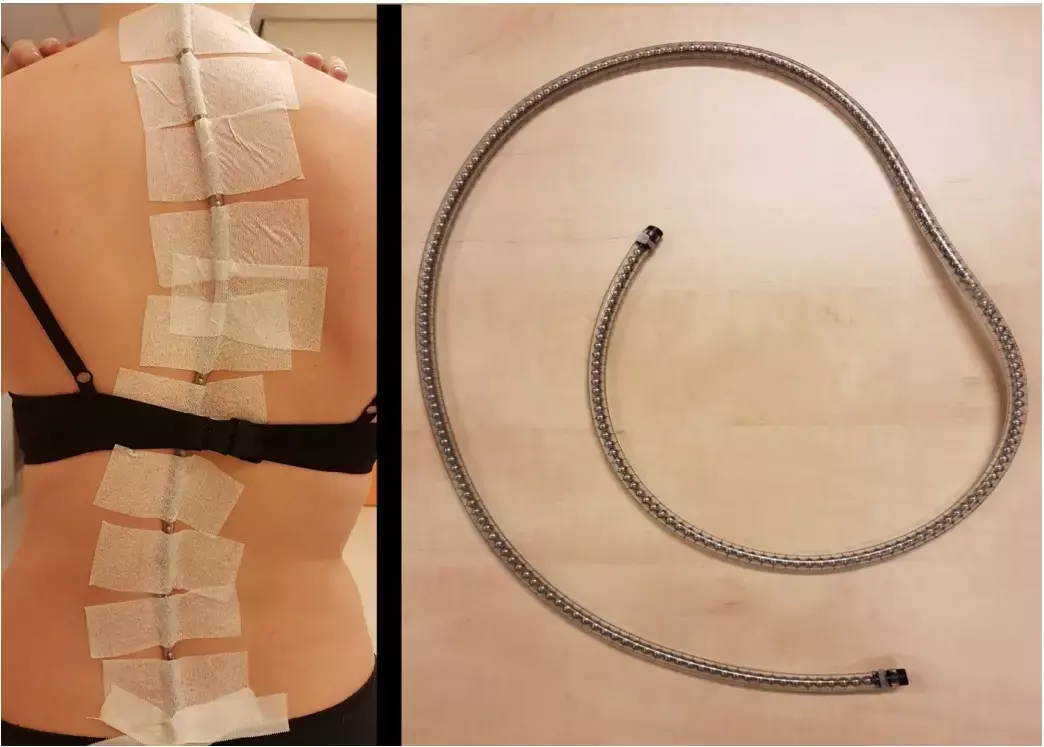
Prior to routine EOS radiograph, a radiographic calibrated metal beads chain (MBC) with metal beads (5 mm in diameter) was taped to the skin on the spinous processes from vertebra C7 to L5 of 120 patients with AIS to calibrate the images. The physician assistant (JB) placed the chain on the skin of all included patients by carefully palpating each individual spinous process to position the chain parallel to the curve before the patient was positioned on the EOS platform in standing position. Subsequently, the biplanar low-dose radiographs of the spine were conducted. Two observers (CP and FW) independently examined the EOS images for eligibility. Only radiographs with the metal beads chain (MBC) positioned accurately over the spinous processes in parallel to the spine were included for analysis. Any differences or uncertainty concerning the inclusion of the radiographs was solved in a consensus meeting.
Spinal lengths were measured from vertebra to vertebra on EOS anteroposterior (AP), lateral view and on the combined 3D EOS view (EOS 3D). These measurements were compared with MBC length measurements. Secondly, intra- and interobserver reliability of length measurements on EOS-images were determined.
Key findings of the study:
• Of the 120 patients fulfilling the inclusion criteria, 50 patients (41.7%) had good parallel placement of the MBC and were included for analysis.
• The mean age of the 50 included patients at time of inclusion was 17.6 years (SD=3.3) with a range from 12 to 29 years.
• Forty-five patients (90%) were female.
• The correlations between EOS and MBC were highest for the 3D length measurements.
• Compared to EOS 3D measurements, the total spinal length was systematically measured 4.3% (mean difference=1.97±1.12 cm) and 1.9% (mean difference=0.86±0.63 cm) smaller on individual EOS two-dimensional (2D) AP and lateral view images, respectively.
• Both intra- and interobserver reliability were excellent for all length measurements on EOS-images.
The authors concluded that – “this study shows a good validity and reliability for spinal length measurements on EOS radiographs. The EOS 3D length measure method is preferred above 2D spinal length measurements on EOS AP or lateral views and can be used for total, thoracic, lumbar or segmental spinal length measurements in AIS patients. When the EOS 3D measure method is not possible, spinal length measurements on EOS 2D (lateral view) could be preferred above measurements on EOS 2D (AP view) when coronal Cobb angle is below 40 degrees. In the future, an automated spinal length measurement system would be helpful in accurate timing of treatment.”
Further reading:
Assessment of spine length in scoliosis patients using EOS imaging: a validity and reliability study
C. M. M. Peeters, G. J. F. J. Bos et al
European Spine Journal (2022) 31:3527–3535
https://doi.org/10.1007/s00586-022-07326-4
MBBS, Dip. Ortho, DNB ortho, MNAMS
Dr Supreeth D R (MBBS, Dip. Ortho, DNB ortho, MNAMS) is a practicing orthopedician with interest in medical research and publishing articles. He completed MBBS from mysore medical college, dip ortho from Trivandrum medical college and sec. DNB from Manipal Hospital, Bengaluru. He has expirence of 7years in the field of orthopedics. He has presented scientific papers & posters in various state, national and international conferences. His interest in writing articles lead the way to join medical dialogues. He can be contacted at editorial@medicaldialogues.in.
Dr Kamal Kant Kohli-MBBS, DTCD- a chest specialist with more than 30 years of practice and a flair for writing clinical articles, Dr Kamal Kant Kohli joined Medical Dialogues as a Chief Editor of Medical News. Besides writing articles, as an editor, he proofreads and verifies all the medical content published on Medical Dialogues including those coming from journals, studies,medical conferences,guidelines etc. Email: drkohli@medicaldialogues.in. Contact no. 011-43720751


