- Home
- Medical news & Guidelines
- Anesthesiology
- Cardiology and CTVS
- Critical Care
- Dentistry
- Dermatology
- Diabetes and Endocrinology
- ENT
- Gastroenterology
- Medicine
- Nephrology
- Neurology
- Obstretics-Gynaecology
- Oncology
- Ophthalmology
- Orthopaedics
- Pediatrics-Neonatology
- Psychiatry
- Pulmonology
- Radiology
- Surgery
- Urology
- Laboratory Medicine
- Diet
- Nursing
- Paramedical
- Physiotherapy
- Health news
- Fact Check
- Bone Health Fact Check
- Brain Health Fact Check
- Cancer Related Fact Check
- Child Care Fact Check
- Dental and oral health fact check
- Diabetes and metabolic health fact check
- Diet and Nutrition Fact Check
- Eye and ENT Care Fact Check
- Fitness fact check
- Gut health fact check
- Heart health fact check
- Kidney health fact check
- Medical education fact check
- Men's health fact check
- Respiratory fact check
- Skin and hair care fact check
- Vaccine and Immunization fact check
- Women's health fact check
- AYUSH
- State News
- Andaman and Nicobar Islands
- Andhra Pradesh
- Arunachal Pradesh
- Assam
- Bihar
- Chandigarh
- Chattisgarh
- Dadra and Nagar Haveli
- Daman and Diu
- Delhi
- Goa
- Gujarat
- Haryana
- Himachal Pradesh
- Jammu & Kashmir
- Jharkhand
- Karnataka
- Kerala
- Ladakh
- Lakshadweep
- Madhya Pradesh
- Maharashtra
- Manipur
- Meghalaya
- Mizoram
- Nagaland
- Odisha
- Puducherry
- Punjab
- Rajasthan
- Sikkim
- Tamil Nadu
- Telangana
- Tripura
- Uttar Pradesh
- Uttrakhand
- West Bengal
- Medical Education
- Industry
Case of Treatment of Traumatic Critical-Sized Tibial Bone Defect with Largest 3D printed cage: a report

Traditional approaches to treat Critical-Sized bone Defects (CSDs) of the foot and ankle are cancellous bone allografts, vascularized autografts, induced membrane or Masquelet technique, and bone transport. These are often inadequate requiring multiple procedures and substantial patient cooperation. Three-dimensional (3D) printing offers a promising solution to the challenge of CSDs.
Lindsey G. Johnson et al reported a case of a 21-year-old woman who sustained an open, severely comminuted, Grade III pilon fracture of the left ankle secondary to a high-speed motor vehicle accident as a restrained driver.
The patient was initially treated at an outside facility with wound debridement, primary closure, and external fixation.
Owing to the sole recommendation of below-the-knee amputation, the patient presented almost 3 months later to Duke University Medical Center, Durham, NC for a second opinion. She presented in external fixation with palpable dorsalispedis and tibialis posterior pulses and intact sensation throughout the foot with complete wound healing and no evidence of infection or contamination.
Radiographic imaging indicated a severely complex, intra-articular comminuted fracture of the distal tibial metadiaphysis with extension into the tibiotalar joint. There was evidence of concomitant mildly comminuted fractures of the distal fibular metadiaphysis with a displaced bone fragment.
Most importantly, there was a CSD of unsalvageable tibial bone measuring 12 cm, limiting traditional surgical reconstruction options.
The patient elected to proceed with limb salvage by arthrodesis of the tibia to the hindfoot using a custom, 3D printed titanium cage (Restor3d, Durham, NC) and an intramedullary rod. The interdisciplinary planning session with mechanical engineers was based on the radiograph and computed tomography of the contralateral limb to construct a custom, 3D printed anatomical template. During the planning session, the foot was repositioned under the tibia until anatomical alignment was obtained. Additional proposed bony resection allowed for a transverse cut of the tibia in both sagittal and anteroposterior planes. The remaining defect, after alignment and revision tibial osteotomy, measured 12 cm in length. Computer modeling was performed to create a cage to fill the resultant defect.
The external fixator was removed under anesthesia without evidence of infection or complication. A 1-month “pin-site holiday” was performed before definitive surgical management, during which the patient was placed in a short leg cast and kept non–weight-bearing. Subsequently, the custom, 3D printed implant was placed in the CSD along with an intramedullary tibiotalocalcaneal (TTC) arthrodesis nail for stabilization of the 3D implant to the surrounding bone.
A standard anterior approach to the ankle and distal tibia was performed. A midline incision was made with deep dissection between the tibialis anterior and extensor halluces longus. An autogenous bone graft was harvested from the left tibia, and an allograft bone (Blackstone Medical Inc; Pioneer Surgical Technology) was transplanted to fill the entire porous lattice structure of the implant to promote osseointegration. A second, lateral incision over the fibula was made, and a small amount of the fibula was osteotomized and a bone mill was morselized. The morselized fibula was placed within the central cavity of the cage and distributed into the pores of the implant.
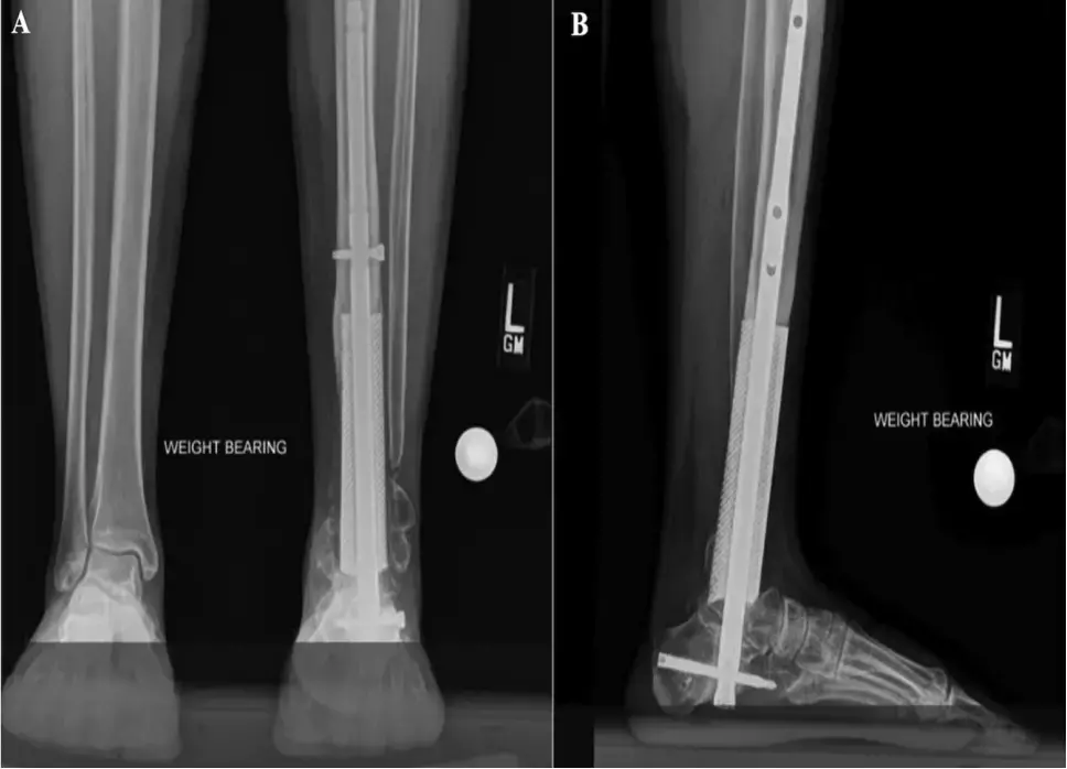
Follow-Up and Outcomes After surgery, the patient remained non–weight-bearing for 6 weeks, followed by a 6-week period of limited weight-bearing in a cast. She was then transitioned to full weight-bearing in a boot brace over the final 6 weeks. Serial radiographs were obtained at 6- to 12-week intervals to monitor for signs of degenerative changes, areas of lucency, or loss of structural integrity.
Radiographic imaging demonstrated successful bony integration of the tibia, talus, and calcaneus into the titanium gyroid lattice. The patient is currently employed in an occupation requiring standing for 8 hours per day and exhibited normal gait biomechanics at the final follow-up.
The authors concluded that – ‘3D printing offers a novel solution to CSDs. To the best of our knowledge, this case report details the largest 3D printed cage, to date, used to treat tibial bone loss. This report describes a unique approach to traumatic limb salvage with favorable patient-reported outcomes and evidence of radiographic fusion at a 3-year follow up.’
Further reading:
Three-Year Follow-Up of a Traumatic Critical-Sized Tibial Bone Defect Treated with a 3D Printed Titanium Cage A Case ReportLindsey G. Johnson, Molly M. Kearney et al JBJS Case Connect 2023;13:e22.00077http://dx.doi.org/10.2106/JBJS.CC.22.00077
MBBS, Dip. Ortho, DNB ortho, MNAMS
Dr Supreeth D R (MBBS, Dip. Ortho, DNB ortho, MNAMS) is a practicing orthopedician with interest in medical research and publishing articles. He completed MBBS from mysore medical college, dip ortho from Trivandrum medical college and sec. DNB from Manipal Hospital, Bengaluru. He has expirence of 7years in the field of orthopedics. He has presented scientific papers & posters in various state, national and international conferences. His interest in writing articles lead the way to join medical dialogues. He can be contacted at editorial@medicaldialogues.in.
Dr Kamal Kant Kohli-MBBS, DTCD- a chest specialist with more than 30 years of practice and a flair for writing clinical articles, Dr Kamal Kant Kohli joined Medical Dialogues as a Chief Editor of Medical News. Besides writing articles, as an editor, he proofreads and verifies all the medical content published on Medical Dialogues including those coming from journals, studies,medical conferences,guidelines etc. Email: drkohli@medicaldialogues.in. Contact no. 011-43720751


