- Home
- Medical news & Guidelines
- Anesthesiology
- Cardiology and CTVS
- Critical Care
- Dentistry
- Dermatology
- Diabetes and Endocrinology
- ENT
- Gastroenterology
- Medicine
- Nephrology
- Neurology
- Obstretics-Gynaecology
- Oncology
- Ophthalmology
- Orthopaedics
- Pediatrics-Neonatology
- Psychiatry
- Pulmonology
- Radiology
- Surgery
- Urology
- Laboratory Medicine
- Diet
- Nursing
- Paramedical
- Physiotherapy
- Health news
- Fact Check
- Bone Health Fact Check
- Brain Health Fact Check
- Cancer Related Fact Check
- Child Care Fact Check
- Dental and oral health fact check
- Diabetes and metabolic health fact check
- Diet and Nutrition Fact Check
- Eye and ENT Care Fact Check
- Fitness fact check
- Gut health fact check
- Heart health fact check
- Kidney health fact check
- Medical education fact check
- Men's health fact check
- Respiratory fact check
- Skin and hair care fact check
- Vaccine and Immunization fact check
- Women's health fact check
- AYUSH
- State News
- Andaman and Nicobar Islands
- Andhra Pradesh
- Arunachal Pradesh
- Assam
- Bihar
- Chandigarh
- Chattisgarh
- Dadra and Nagar Haveli
- Daman and Diu
- Delhi
- Goa
- Gujarat
- Haryana
- Himachal Pradesh
- Jammu & Kashmir
- Jharkhand
- Karnataka
- Kerala
- Ladakh
- Lakshadweep
- Madhya Pradesh
- Maharashtra
- Manipur
- Meghalaya
- Mizoram
- Nagaland
- Odisha
- Puducherry
- Punjab
- Rajasthan
- Sikkim
- Tamil Nadu
- Telangana
- Tripura
- Uttar Pradesh
- Uttrakhand
- West Bengal
- Medical Education
- Industry
Endoscopic Partial Proximal Hamstring Repair: technical note for safe and effective surgical treatment

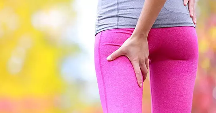
The contemporary treatment of hamstring avulsions has been evolving, as more patients are being identified as having persistently symptomatic partial hamstring tears recalcitrant to nonoperative treatment. The endoscopic hamstring repair allows surgeons improved visualization of the footprint, as well as safe dissection of the sciatic nerve.
B. Capurro et al has described an endoscopic repair technique in an article published in “Arthroscopy Techniques.” This technique article provides a step-by-step technical note to allow for safe and effective surgical treatment of partial hamstring tears.
The limb is positioned such that the hip is extended and the knee flexed, which relaxes both the gluteus maximus muscle and the sciatic nerve. The endoscopic portals are created along the gluteal crease in the event that an open incision is required. The medial portal is located just medial to the lateral border of the tuberosity, and 2 cm distal to the distal border along the gluteal fold. The lateral portal is then made, 3-4 cm lateral to the medial portal, also within the gluteal fold. The portal is made under spinal needle localization to directly visualize the instrument to avoid injury to the sciatic nerve.
A full radius arthroscopic shaver is placed through the lateral portal, and an ischial bursectomy is performed to improve visualization and clear the subgluteal space. After a thorough bursectomy, the next step is to identify the hamstring tendon tear. Use the tip of the shaver or switching stick to palpate the tendon footprint against the ischium to identify the defect. The torn tendon is ballotable relative to the intact tendon. Once the defect has been identified, a radio frequency ablater (RFA) can be used to open the sheath longitudinally to localize the avulsion. Before anchor placement and suture retrieval can be performed, the sciatic nerve must be identified and protected.
The authors’ preference is to use the non-toothed shaver to dissect to the sciatic nerve to avoid inadvertent injury or bleeding from nearby vascular structures. After developing the plane between the hamstring and gluteus maximus, the RFA is used to clear off any soft tissue on the ischium. Next, a 5.5-mm cylindrical burr is used to decorticate the footprint to create a bleeding bed of bone, as well as augment the biologic healing response. Hamstring Repair After punching and tapping, a triple-loaded 5.5-mm Peek AlphaVent anchor is placed at the footprint through a percutaneous portal using a spinal needle. All 6 suture limbs are initially pulled out of the percutaneous portal for suture management.
An 8.0 x 90 mm cannula is placed through the lateral portal, and a tissue penetrator is passed from lateral to medial through the tendon stump. This trajectory aims away from the sciatic nerve, and, therefore, is safer than a medial to lateral direction. The passing suture is used to shuttle the first limb from the anchor through the tendon. This step is repeated for the matching limb of the suture, creating a horizontal mattress construct. This step is repeated until all 6 suture limbs are passed through the tendon stump. Ideally, there is about 0.5 cm of tendon between each limb of a single mattress, and 1 cm between each mattress. Next, the arthroscope is switched to the lateral portal, and the tissue penetrating device is used to retrieve sutures from the medial aspect of the hamstring tear. The arthroscope is placed back into the medial portal, and the sutures are tied using alternating half hitches on alternating posts in a mattress configuration for anatomic hamstring repair. Following repair, meticulous hemostasis is achieved to prevent hematoma formation, which could result in sciatic nerve compression.
The author also mentions the advantages of this technique as mentioned below:
• Minimally invasive, smaller surgical incisions decrease the risk of infection and wound complications.
• Clearer visualization of tendon pathology, particularly partial tears.
• After medial portal is made, lateral portal is made under direct visualization, avoiding iatrogenic neurovascular injury.
• Minimal blood loss.
• Patient positioned in the prone position with the knee partially flexed helps to avoid damage to the neurovascular structures.
• Limits prolonged retraction of the gluteus maximus.
Further reading:
Endoscopic Partial Proximal Hamstring Repair
B. CAPURRO ET AL
Arthroscopy Techniques, Vol 12, No 7 (July), 2023: pp e1075-e1081
https://doi.org/10.1016/j.eats.2023.02.045
MBBS, Dip. Ortho, DNB ortho, MNAMS
Dr Supreeth D R (MBBS, Dip. Ortho, DNB ortho, MNAMS) is a practicing orthopedician with interest in medical research and publishing articles. He completed MBBS from mysore medical college, dip ortho from Trivandrum medical college and sec. DNB from Manipal Hospital, Bengaluru. He has expirence of 7years in the field of orthopedics. He has presented scientific papers & posters in various state, national and international conferences. His interest in writing articles lead the way to join medical dialogues. He can be contacted at editorial@medicaldialogues.in.
Dr Kamal Kant Kohli-MBBS, DTCD- a chest specialist with more than 30 years of practice and a flair for writing clinical articles, Dr Kamal Kant Kohli joined Medical Dialogues as a Chief Editor of Medical News. Besides writing articles, as an editor, he proofreads and verifies all the medical content published on Medical Dialogues including those coming from journals, studies,medical conferences,guidelines etc. Email: drkohli@medicaldialogues.in. Contact no. 011-43720751


