- Home
- Medical news & Guidelines
- Anesthesiology
- Cardiology and CTVS
- Critical Care
- Dentistry
- Dermatology
- Diabetes and Endocrinology
- ENT
- Gastroenterology
- Medicine
- Nephrology
- Neurology
- Obstretics-Gynaecology
- Oncology
- Ophthalmology
- Orthopaedics
- Pediatrics-Neonatology
- Psychiatry
- Pulmonology
- Radiology
- Surgery
- Urology
- Laboratory Medicine
- Diet
- Nursing
- Paramedical
- Physiotherapy
- Health news
- Fact Check
- Bone Health Fact Check
- Brain Health Fact Check
- Cancer Related Fact Check
- Child Care Fact Check
- Dental and oral health fact check
- Diabetes and metabolic health fact check
- Diet and Nutrition Fact Check
- Eye and ENT Care Fact Check
- Fitness fact check
- Gut health fact check
- Heart health fact check
- Kidney health fact check
- Medical education fact check
- Men's health fact check
- Respiratory fact check
- Skin and hair care fact check
- Vaccine and Immunization fact check
- Women's health fact check
- AYUSH
- State News
- Andaman and Nicobar Islands
- Andhra Pradesh
- Arunachal Pradesh
- Assam
- Bihar
- Chandigarh
- Chattisgarh
- Dadra and Nagar Haveli
- Daman and Diu
- Delhi
- Goa
- Gujarat
- Haryana
- Himachal Pradesh
- Jammu & Kashmir
- Jharkhand
- Karnataka
- Kerala
- Ladakh
- Lakshadweep
- Madhya Pradesh
- Maharashtra
- Manipur
- Meghalaya
- Mizoram
- Nagaland
- Odisha
- Puducherry
- Punjab
- Rajasthan
- Sikkim
- Tamil Nadu
- Telangana
- Tripura
- Uttar Pradesh
- Uttrakhand
- West Bengal
- Medical Education
- Industry
Imaging of ballistic wounds may predict retained bullet composition and MRI safety

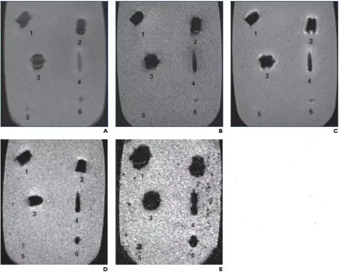
CAPTION
Scout (A), T1-weighted spin-echo (SE) (B), T2-weighted SE (C), T2-weighted gradient-recalled echo (GRE) (TR/TE, 500/10; D), and T2-weighted GRE (TR/TE, 700/30; E) MR images show jacket hollow point .45 automatic Colt pistol bullet (Corbon) (1), solid lead .45 Long Colt bullet (Winchester) (2), full metal jacket (FMJ) automatic Colt pistol bullet (Winchester) (3), 5.56-mm FMJ bullet (Federal Ammunition) (4), #7 lead shotgun pellet (Winchester) (5), and 5-mm lead air gun pellet (Sheridan) (6). On all sequences, metallic artifact is minimal. Although metallic artifact increases or blooms with increased TR/TE in GRE images (D and E), amount of surrounding distortion is still minimal.
CREDIT
American Roentgen Ray Society (ARRS), American Journal of Roentgenology (AJR)
Atlanta, GA: Magntic Resonance Imaging is frequently denied to patients with ballistic embedded fragments because without shell casing it is not possible to determine the bullet composition. Now, a recent study in the American Journal of Roentgenology has found that CT and radiography can be used for the identification of nonferromagnetic projectiles that are safe for MRI. The researchers also presented an algorithm for the determination of the triage of patients with retained bullets.
The purpose of this article by Arthur J. Fountain, Emory University, Atlanta, GA, and colleagues aimed to determined whether the radiographic and CT appearance of ballistic projectiles predicts their composition. They also characterized the translational, rotational, and temperature effects of a 1.5-T MRI magnetic field on representative bullets.
For the purpose, commercially available handgun and shotgun ammunition representing projectiles commonly encountered in a clinical setting was fired into ballistic gelatin as a surrogate for human tissue. Radiographs and CT images of these gelatin blocks were obtained. Using T1- and T2-weighted sequences, MR images of unfired bullets suspended in gelatin blocks were also obtained. Magnetic attractive force, rotational torque, and heating effects of unfired bullets were assessed at 1.5 T.
Key findings of the study include:
- Fired bullets were separated into ferromagnetic and nonferromagnetic groups based on the presence of a debris trail and deformation of the primary projectile in the gelatin blocks.
- Whereas ferromagnetic bullets showed mild torque forces and marked imaging artifacts at 1.5 T, nonferromagnetic bullets did not have these effects.
- Heating above the Food and Drug Administration limit of 2°C was not observed in any of the projectiles tested.
"We found that radiography and CT can be used to identify nonferromagnetic projectiles that are safe for MRI," concluded the authors. "We also present an algorithm for determining the triage of patients with retained bullets."
"Nonferromagnetic ballistic projectiles do not undergo movement or heating during MRI, and the imaging modality can be performed when medically necessary without undue risk and with limited artifact susceptibility on the resulting images, even when the projectile is in or near a vital structure," the authors concluded.
The study, "Imaging Appearance of Ballistic Wounds Predicts Bullet Composition: Implications for MRI Safety," is published in the American Journal of Roentgenology.
DOI: https://www.ajronline.org/doi/abs/10.2214/AJR.20.23648
Dr Kamal Kant Kohli-MBBS, DTCD- a chest specialist with more than 30 years of practice and a flair for writing clinical articles, Dr Kamal Kant Kohli joined Medical Dialogues as a Chief Editor of Medical News. Besides writing articles, as an editor, he proofreads and verifies all the medical content published on Medical Dialogues including those coming from journals, studies,medical conferences,guidelines etc. Email: drkohli@medicaldialogues.in. Contact no. 011-43720751


