- Home
- Medical news & Guidelines
- Anesthesiology
- Cardiology and CTVS
- Critical Care
- Dentistry
- Dermatology
- Diabetes and Endocrinology
- ENT
- Gastroenterology
- Medicine
- Nephrology
- Neurology
- Obstretics-Gynaecology
- Oncology
- Ophthalmology
- Orthopaedics
- Pediatrics-Neonatology
- Psychiatry
- Pulmonology
- Radiology
- Surgery
- Urology
- Laboratory Medicine
- Diet
- Nursing
- Paramedical
- Physiotherapy
- Health news
- Fact Check
- Bone Health Fact Check
- Brain Health Fact Check
- Cancer Related Fact Check
- Child Care Fact Check
- Dental and oral health fact check
- Diabetes and metabolic health fact check
- Diet and Nutrition Fact Check
- Eye and ENT Care Fact Check
- Fitness fact check
- Gut health fact check
- Heart health fact check
- Kidney health fact check
- Medical education fact check
- Men's health fact check
- Respiratory fact check
- Skin and hair care fact check
- Vaccine and Immunization fact check
- Women's health fact check
- AYUSH
- State News
- Andaman and Nicobar Islands
- Andhra Pradesh
- Arunachal Pradesh
- Assam
- Bihar
- Chandigarh
- Chattisgarh
- Dadra and Nagar Haveli
- Daman and Diu
- Delhi
- Goa
- Gujarat
- Haryana
- Himachal Pradesh
- Jammu & Kashmir
- Jharkhand
- Karnataka
- Kerala
- Ladakh
- Lakshadweep
- Madhya Pradesh
- Maharashtra
- Manipur
- Meghalaya
- Mizoram
- Nagaland
- Odisha
- Puducherry
- Punjab
- Rajasthan
- Sikkim
- Tamil Nadu
- Telangana
- Tripura
- Uttar Pradesh
- Uttrakhand
- West Bengal
- Medical Education
- Industry
MRI may detect Concussion-Linked CTE while Patients are still alive

Chronic traumatic encephalopathy (CTE), a neurodegenerative tauopathy, cannot be diagnosed during life at the moment. Atrophy patterns on magnetic resonance imaging may be an effective in vivo biomarker of CTE, but they have yet to be identified. Neurodegeneration mechanisms in CTE are unknown.
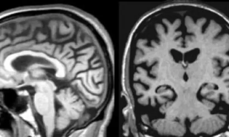
Magnetic Resonance Imaging may be able to detect chronic traumatic encephalopathy (CTE) while people are still alive finds a new study. As of now Chronic traumatic encephalopathy (CTE) which is a devastating concussion-linked brain condition can only be diagnosed after death via autopsy.
This study was conducted by Michael L. Alosco and team with the objective to Describe the macrostructural magnetic resonance imaging features of brain donors with autopsy-confirmed CTE, as well as the relationship between hyperphosphorylated tau (p-tau) and atrophy on magnetic resonance imaging. The findings of this study were published in BMC - Alzheimer's Research & Therapy.
In this study magnetic resonance imaging scans were obtained through medical record requests for 55 deceased symptomatic men with autopsy-confirmed CTE and 31 men (n = 11 deceased) with normal cognition at the time of the scan, all of whom were over 60 years old. Three neuroradiologists assessed regional atrophy and microvascular disease (0 [none]–4 [severe]), microbleeds, and the presence of cavum septum pellucidum. At autopsy, neuropathologists used semi-quantitative scales to assess tau severity and atrophy.
Results stated as follow:
1. When compared to unimpaired males, donors with CTE (45/55=stage III/IV) had greater atrophy of the orbital-frontal (mean diff.=1.29), dorsolateral frontal (mean diff.=1.31), superior frontal (mean diff.=1.05), anterior temporal (mean diff.=1.57), and medial temporal lobes (mean diff.=1.60), and larger lateral (mean diff.=1.7).
2. No effects were observed for posterior atrophy or microvascular disease. Donors with CTE were more likely to have a cavum septum pellucidum (OR = 6.7, p 0.05).
3. Greater tau severity across 14 regions in CTE donors corresponded to greater atrophy on magnetic resonance imaging (beta = 0.68, p 0.01).
In conclusion, when compared to same-age men with NC, cognitively symptomatic male brain donors with autopsy-confirmed CTE had more severe visually rated frontal, temporal, and hippocampal atrophy, as well as an increased risk of having a CSP on antemortem MRI scans. Furthermore, more severe p-tau pathology was linked to higher MRI atrophy ratings. These findings, if validated by prospective clinical-pathological correlation studies, support the use of structural MRI as a valuable tool for supporting a CTE diagnosis during life.
Reference:
Alosco, M.L., Mian, A.Z., Buch, K. et al. Structural MRI profiles and tau correlates of atrophy in autopsy-confirmed CTE. Alz Res Therapy 13, 193 (2021). https://doi.org/10.1186/s13195-021-00928-y
Medical Dialogues consists of a team of passionate medical/scientific writers, led by doctors and healthcare researchers. Our team efforts to bring you updated and timely news about the important happenings of the medical and healthcare sector. Our editorial team can be reached at editorial@medicaldialogues.in.
Dr Kamal Kant Kohli-MBBS, DTCD- a chest specialist with more than 30 years of practice and a flair for writing clinical articles, Dr Kamal Kant Kohli joined Medical Dialogues as a Chief Editor of Medical News. Besides writing articles, as an editor, he proofreads and verifies all the medical content published on Medical Dialogues including those coming from journals, studies,medical conferences,guidelines etc. Email: drkohli@medicaldialogues.in. Contact no. 011-43720751


