- Home
- Medical news & Guidelines
- Anesthesiology
- Cardiology and CTVS
- Critical Care
- Dentistry
- Dermatology
- Diabetes and Endocrinology
- ENT
- Gastroenterology
- Medicine
- Nephrology
- Neurology
- Obstretics-Gynaecology
- Oncology
- Ophthalmology
- Orthopaedics
- Pediatrics-Neonatology
- Psychiatry
- Pulmonology
- Radiology
- Surgery
- Urology
- Laboratory Medicine
- Diet
- Nursing
- Paramedical
- Physiotherapy
- Health news
- Fact Check
- Bone Health Fact Check
- Brain Health Fact Check
- Cancer Related Fact Check
- Child Care Fact Check
- Dental and oral health fact check
- Diabetes and metabolic health fact check
- Diet and Nutrition Fact Check
- Eye and ENT Care Fact Check
- Fitness fact check
- Gut health fact check
- Heart health fact check
- Kidney health fact check
- Medical education fact check
- Men's health fact check
- Respiratory fact check
- Skin and hair care fact check
- Vaccine and Immunization fact check
- Women's health fact check
- AYUSH
- State News
- Andaman and Nicobar Islands
- Andhra Pradesh
- Arunachal Pradesh
- Assam
- Bihar
- Chandigarh
- Chattisgarh
- Dadra and Nagar Haveli
- Daman and Diu
- Delhi
- Goa
- Gujarat
- Haryana
- Himachal Pradesh
- Jammu & Kashmir
- Jharkhand
- Karnataka
- Kerala
- Ladakh
- Lakshadweep
- Madhya Pradesh
- Maharashtra
- Manipur
- Meghalaya
- Mizoram
- Nagaland
- Odisha
- Puducherry
- Punjab
- Rajasthan
- Sikkim
- Tamil Nadu
- Telangana
- Tripura
- Uttar Pradesh
- Uttrakhand
- West Bengal
- Medical Education
- Industry
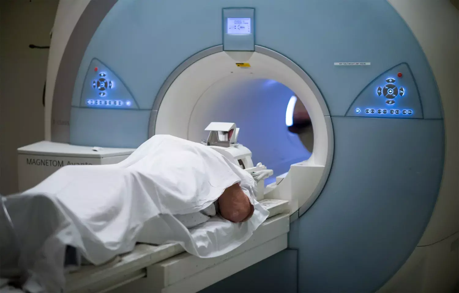
New cardiac MRI protocol provides complete heart study in less than a minute: Study

Spain: A new MRI technique allows complete cardiac examination in less than a minute, according to a recent study in the journal JACC: Cardiovascular Imaging.
The technique, called SENSE by static outer-volume subtraction (ESSOS), is developed by Royal Philips and the Spanish National Center for Cardiovascular Research, and is based on the fact that -- except for the heart -- a patient's chest anatomy remains stationary during a breath-hold exam.
Cardiac magnetic resonance is the reference tool for cardiac imaging but is time-consuming. SandraGómez-Talavera, Department of Cardiology, IIS-Hospital Fundacion Jiménez Díaz, Madrid, Spain, and colleague, therefore, aimed to clinically validate a novel 3-dimensional (3D) ultrafast cardiac magnetic resonance (CMR) protocol including cine (anatomy and function) and late gadolinium enhancement (LGE), each in a single breath-hold.
In a prospective study of 107 patients, the researchers compared protocol comprising isotropic 3D cine (Enhanced sensitivity encoding [SENSE] by Static Outer volume Subtraction [ESSOS]) and isotropic 3D LGE sequences with a standard cine+LGE protocol. Left ventricular (LV) mass, volumes, and LV and right ventricular (RV) ejection fraction (LVEF, RVEF) were assessed by 3D ESSOS and 2D cine CMR. LGE (% LV) was assessed using 3D and 2D sequences.
The study revealed the following findings:
- Three-dimensional and LGE acquisitions lasted 24 and 22 s, respectively.
- Three-dimensional and LGE images were of good quality and allowed quantification in all cases.
- Mean LVEF by 3D and 2D CMR were 51 ± 12% and 52 ± 12%, respectively, with excellent intermethod agreement (intraclass correlation coefficient [ICC]: 0.96) and insignificant bias.
- Mean RVEF 3D and 2D CMR were 60.4 ± 5.4% and 59.7 ± 5.2%, respectively, with acceptable intermethod agreement (ICC: 0.73) and insignificant bias.
- Both 2D and 3D LGE showed excellent agreement, and intraobserver and interobserver agreement were excellent for 3D LGE.
Based on the findings the team concluded that ESSOS single breath-hold 3D CMR allows accurate assessment of heart anatomy and function.
"Combining ESSOS with 3D LGE allows complete cardiac examination in <1 min of acquisition time. This protocol expands the indication for CMR, reduces costs, and increases patient comfort," they wrote.
Reference:
The study titled, "Clinical Validation of a 3-Dimensional Ultrafast Cardiac Magnetic Resonance Protocol Including Single Breath-Hold 3-Dimensional Sequences," is published in the journal JACC: Cardiovascular Imaging.
DOI: https://www.sciencedirect.com/science/article/pii/S1936878X21002849
Dr Kamal Kant Kohli-MBBS, DTCD- a chest specialist with more than 30 years of practice and a flair for writing clinical articles, Dr Kamal Kant Kohli joined Medical Dialogues as a Chief Editor of Medical News. Besides writing articles, as an editor, he proofreads and verifies all the medical content published on Medical Dialogues including those coming from journals, studies,medical conferences,guidelines etc. Email: drkohli@medicaldialogues.in. Contact no. 011-43720751


